. 2014 Jan;21(1):146-60.
doi: 10.1038/cdd.2013.141. Epub 2013 Oct 18.

p34 is a novel regulator of the oncogenic behavior of NEDD4-1 and PTEN

Affiliations

p34 is a novel regulator of the oncogenic behavior of NEDD4-1 and PTEN

S-W Hong et al. Cell Death Differ. 2014 Jan.

Abstract

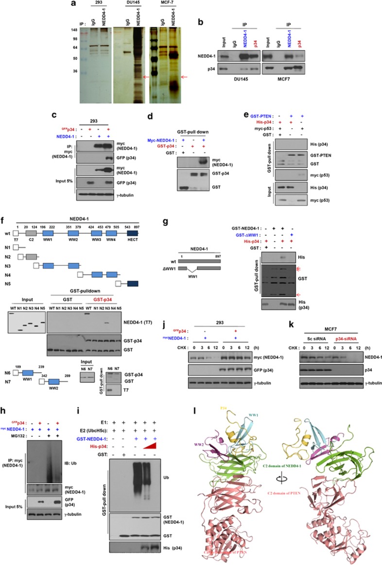
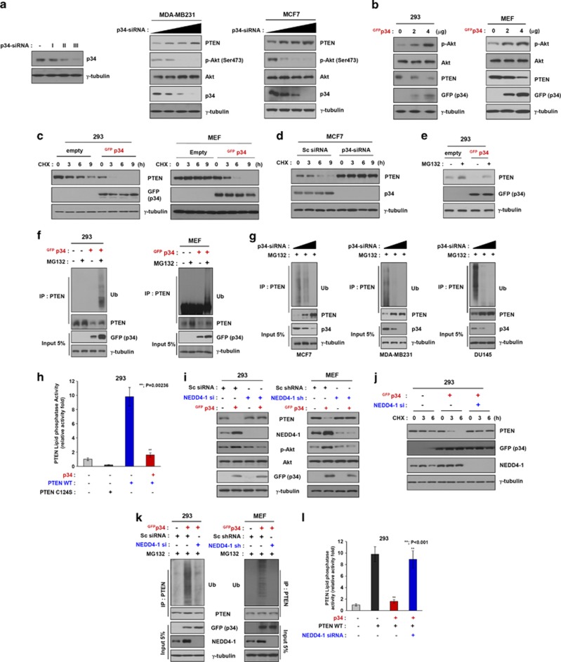
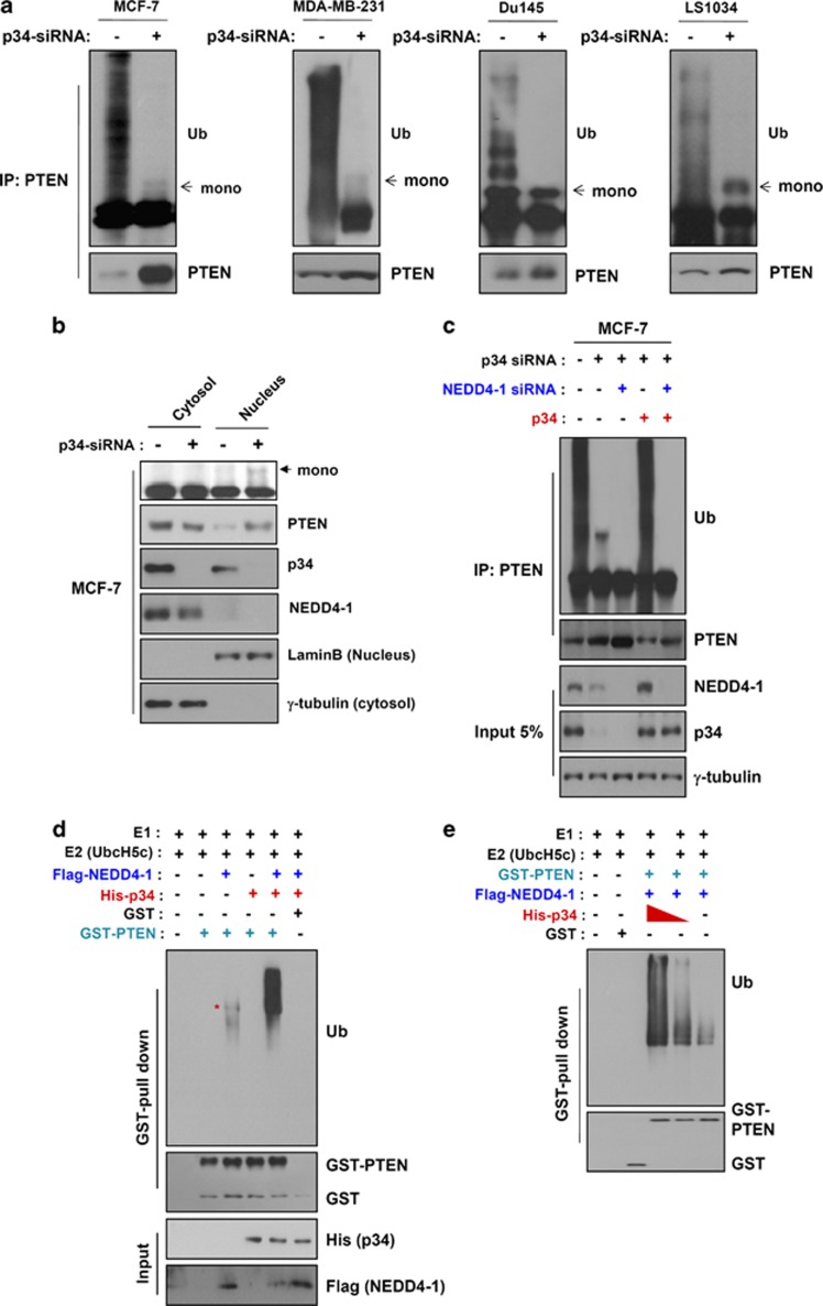
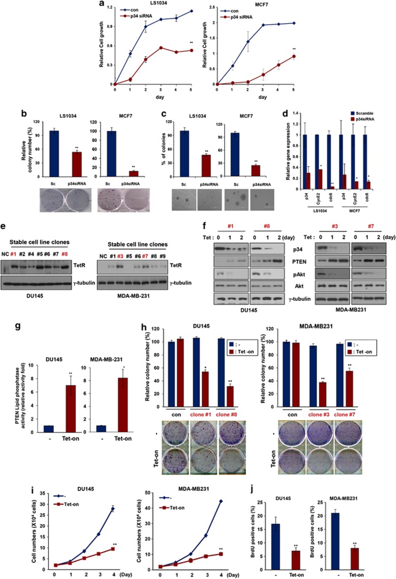
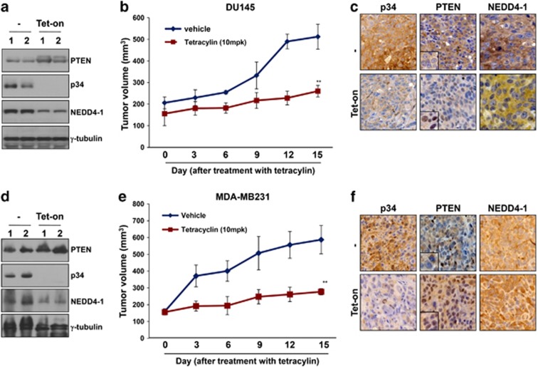
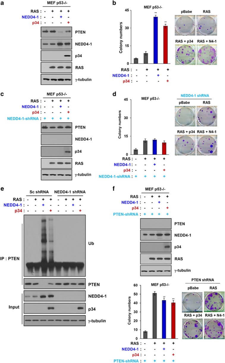
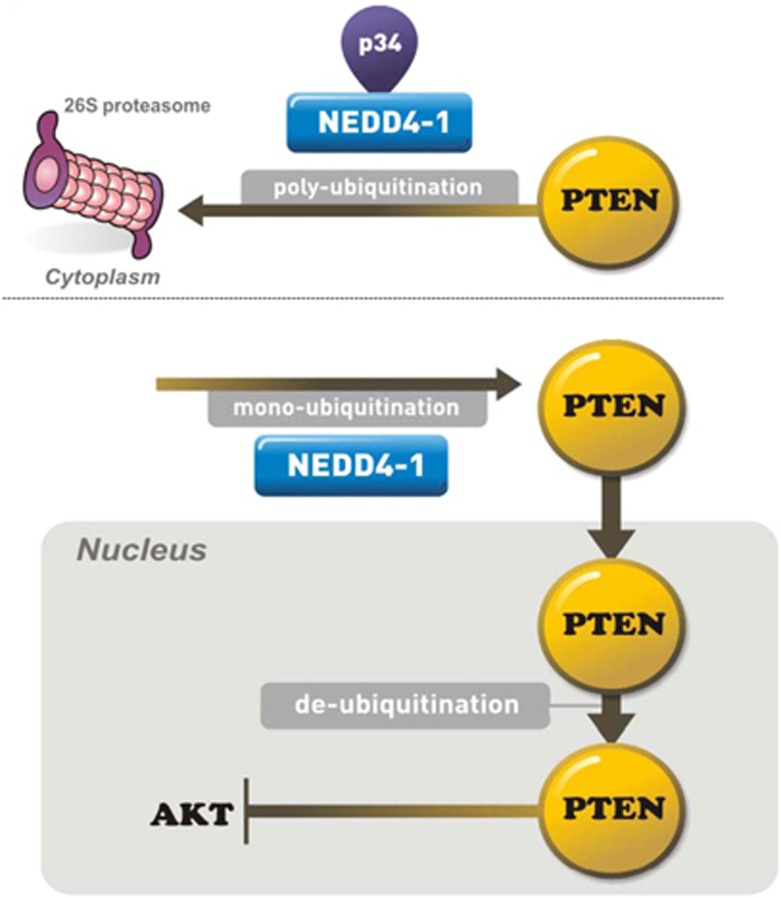
PTEN is one of the most frequently mutated or deleted tumor suppressors in human cancers. NEDD4-1 was recently identified as the E3 ubiquitin ligase for PTEN; however, a number of important questions remain regarding the role of ubiquitination in regulating PTEN function and the mechanisms by which PTEN ubiquitination is regulated. In the present study, we demonstrated that p34, which was identified as a binding partner of NEDD4-1, controls PTEN ubiquitination by regulating NEDD4-1 protein stability. p34 interacts with the WW1 domain of NEDD4-1, an interaction that enhances NEDD4-1 stability. Expression of p34 promotes PTEN poly-ubiquitination, leading to PTEN protein degradation, whereas p34 knockdown results in PTEN mono-ubiquitination. Notably, an inverse correlation between PTEN and p34/NEDD4-1 levels was confirmed in tumor samples from colon cancer patients. Thus, p34 acts as a key regulator of the oncogenic behavior of NEDD4-1 and PTEN.


Figures

Figure 1
Figure 1
p34 interacts with NEDD4-1 and enhances NEDD4-1 stability. (a) p34 is expressed in 293, DU145, MCF7, and LS1034 cancer cells (red arrow). Whole-cell lysates were immunoprecipitated using an anti-NEDD4-1 antibody, resolved using SDS-PAGE and analyzed with silver staining. Rabbit IgG was used as a negative immunoprecipitation control. (b) NEDD4-1 interacts with endogenous p34 in intact cells. DU145, MCF7, LS1034, and MDA-MB-231 cell lysates were immunoprecipitated using anti-NEDD4-1 or anti-p34 antibodies and analyzed using western blotting. (c) Exogenously expressed p34 and NEDD4-1 interact. Whole-cell lysates of 293 cells that had been transfected with GFP-p34 and/or NEDD4-1-Myc were analyzed using immunoblotting. (d) NEDD4-1 binds to p34 in vitro. GST-p34 recombinant proteins were incubated with Myc-NEDD4-1-transfected 293 cell lysates, followed by the addition of GST-Sepharose beads. The bead-bound proteins were probed using immunoblotting with antibodies that had been raised against p34 and NEDD4-1. (e) p34 does not directly bind to PTEN. p34 and PTEN binding was analyzed by incubating purified recombinant GST-PTEN with Myc-NEDD4-1. The interaction of p34 with NEDD4-1 was confirmed using His-p34-expressing cells. GST was used as a negative control for the GST pull-down binding assays. p53 was used as a positive control for the PTEN-binding assays. (f) p34 interacts with the WW1 domain of NEDD4-1. The cells were transfected with various NEDD4-1 deletion mutants were the lacking C2, WWs, and the homologous to the E6-AP carboxyl terminus (HECT) domains (NEDD4-1-N1); WWs and the HECT domains (NEDD4-1-N2); C2, WW3, WW4, and the HECT domains (NEDD4-1-N3); C2, WW1, WW2, and the HECT domains (NEDD4-1-N4); or C2 and all WW domains (NEDD4-1-N5). Twenty-four hours later, the cell lysates were incubated with GST-p34 or GST proteins, and the p34-binding domain of NEDD4-1 was analyzed using an in vitro GST pull-down assay. (g) p34 does not bind to a NEDD4-1 WW1 deletion mutant. NEDD4-1-WT or WW1 deletion mutant proteins were incubated with lysates from His-p34-expressing cells, and the binding regions of NEDD4-1 and p34 were analyzed using a GST pull-down assay. The arrows indicate GST, GST-NEDD4-1-WT, and the GST-WW1 deletion mutant. (h) NEDD4-1 auto-ubiquitination is decreased in intact cells in the presence of p34. 293 cells were transfected with NEDD4-1 and/or p34 and then treated with MG132 (25 μM) for 6 h. After harvesting the cells, anti-Myc immunoprecipitates of the cell lysates were analyzed for NEDD4-1 auto-ubiquitination. (i) NEDD4-1 auto-ubiquitination is decreased in a cell-free system in the presence of p34. GST-NEDD4-1 proteins were incubated with ATP, E1, E2, and His-p34, and auto-ubiquitination was measured. (j and k) p34 regulates NEDD4-1 protein levels. 293 and MCF7 cells were transfected with either a p34 expression plasmid for 24 h (j) or with siRNA against p34 for 48 h (k). The cells were treated with cycloheximide (50 μg/ml) at the indicated time points, followed using western blot analyses to determine the NEDD4-1 protein levels. p34 overexpression increases NEDD4-1 levels, whereas siRNA-mediated p34 knockdown decreases NEDD4-1 levels. (l) Ribbon diagram of the docking model structure of the p34/NEDD4-1/PTEN complex. The predicted mode of interactions between p34/NEDD4-1 and PTEN was based on protein–protein docking. p34 is depicted by a yellow ribbon diagram, NEDD4-1 is shown as a green ribbon diagram, and PTEN is depicted as a light red ribbon diagram. Two views from a 90-degree rotation about the vertical axis are shown
Figure 2
Figure 2
p34 regulates NEDD4-1-mediated PTEN poly-ubiquitination. (a) Western blot analysis of MDA-MB-231 and MCF7 cell lines that had been transfected with p34-siRNA revealed that p34-siRNA induces a dose-dependent increase in PTEN levels and a decrease in phospho-AKT (p-AKT). (b) A Western blot analysis of 293 and MEF cells that had been transfected with GFP-tagged p34 revealed that PTEN levels are decreased and p-AKT is increased with increasing doses of exogenously expressed p34. (c and d) The half-life of the PTEN protein is decreased (in 293 cells and MEFs) by p34 overexpression and increased (in MCF7 cells) by siRNA-mediated p34 knockdown. (e) Western blot analysis of PTEN in 293 cells that had been transfected with p34 and incubated with or without MG132 revealed that PTEN is decreased in MG132-treated cells exogenously expressing p34. (f) Ubiquitination assays performed in intact 293 cells (left panel) and MEFs (right panel) that had been transfected with GFP-p34 and incubated with or without MG132 revealed that the poly-ubiquitinated form of PTEN appears following the exogenous expression of p34. (g) Ubiquitination assays performed in intact MCF7, MDA-MB-231, and DU145 cells that were transfected with p34-siRNA for 24 h and incubated with or without MG132 revealed that p34-siRNA induces a dose-dependent decrease in PTEN poly-ubiquitination in these cancer cell lines. (h) p34-mediated poly-ubiquitination of PTEN inhibits PTEN phosphatase activity. The lipid phosphatase activity of PTEN in cells that had been transfected with wild-type PTEN and/or p34 for 24 h was determined by measuring the dephosphorylating activity of immunoprecipitated PTEN. The data represent the means±S.D.'s of at least three independent experiments (**P<0.001). (i) Western blot analysis of PTEN in wild-type and NEDD4-1-knockdown cells that had been transfected with a p34 expression plasmid revealed that PTEN expression is increased by p34 expression in a wild-type NEDD4-1 background but not in NEDD4-1-knockdown cells. (j) An analysis of the PTEN protein half-life in 293 cells that had been transfected with a p34 expression plasmid and NEDD4-1 siRNA revealed that PTEN stability is enhanced by NEDD4-1 knockdown in the context of p34 expression. (k) Intact-cell ubiquitination assays in wild-type and NEDD4-1-knockdown cells that were transfected with a p34 expression plasmid revealed that PTEN poly-ubiquitination is detectable in a wild-type NEDD4-1 background but not in NEDD4-1-knockdown cells. (l) The p34-mediated reductions in the lipid phosphatase activity of PTEN were attenuated in NEDD4-1 siRNA-transfected cells compared with control cells. The data represent the means±S.D.'s of at least three independent experiments (**P<0.001)
Figure 3
Figure 3
p34 promotes switching between NEDD4-1-mediated PTEN mono- and poly-ubiquitination. (a) Intact-cell ubiquitination assays revealed that PTEN is mono-ubiquitinated in p34-knockdown cells (MCF7, MDA-MB-231, DU145, and LS1034). (b) PTEN is localized to the nuclei of p34-knockdown cells. MCF7 cells were transfected with p34-siRNA for 24 h, fractionated into nuclear and cytosolic compartments, and analyzed for PTEN expression using western blot analyses. (c) p34 expression levels dictate the switch from PTEN poly-ubiquitination to mono-ubiquitination. PTEN ubiquitination in intact cells is significantly decreased by p34 and NEDD4-1 knockdown. The cells were transfected with p34-siRNA, NEDD4-1-siRNA, and/or p34 for 24 h. PTEN ubiquitination was determined using western blot analyses of immunoprecipitated PTEN-Ub adducts. (d) Cell-free ubiquitination assays of GST-PTEN that had been incubated with NEDD4-1 in the presence or absence of p34 revealed that PTEN is mono-ubiquitinated in the presence of NEDD4-1 alone and poly-ubiquitinated in the presence of both NEDD4-1 and p34. (e) PTEN poly-ubiquitination in cell-free assays decreases with decreasing doses of p34
Figure 4
Figure 4
p34 regulates cancer cell proliferation. (a) Knockdown of p34 inhibits cell proliferation in the LS1034 (left panel) and MCF7 (right panel) cell lines. The data represent the means±S.D.'s of at least three independent experiments (**P<0.01). (b) Colony-forming assays confirmed that cell proliferation is inhibited in p34-siRNA-transfected LS1034 and MCF7 cell lines. Representative photos are shown in the bottom panels. The data represent the means±S.D.'s of at least three independent experiments (**P<0.01). (c) PTEN suppresses anchorage-independent growth in p34-siRNA-transfected LS1034 and MCF7 cell lines. All soft-agar assays were performed in triplicate. The data represent the means±S.D.'s of at least three independent experiments (**P<0.01). (d) PTEN expression levels were determined using quantitative RT-PCR in cells that had been transfected with p34-siRNA or scrambled siRNA. The data represent the means±S.D.'s of at least three independent experiments (*P<0.05, **P<0.01). (e and f) Cells that had been stably transfected with Tet-On inducible p34-shRNA were treated with tetracycline (Tet; 1 μg/ml) to inhibit p34 expression. PTEN, p34, p-Akt, and NEDD4-1 expressions were analyzed using western blot analysis. γ-tubulin was used as an internal loading control. (g) PTEN lipid phosphatase activity was increased in tetracycline-inducible, stable p34-knockdown cell lines. The data represent the means±S.D.'s of at least three independent experiments (*P<0.05, **P<0.01). (h) Colony-forming assays confirmed that cell proliferation is inhibited in cell lines that have been stably transfected with tetracycline-inducible p34-shRNA. The data represent the means±S.D.'s of at least three independent experiments (*P<0.05, **P<0.01). (i) The growth of MDA-MB-231 and DU145 cells is inhibited in media containing tetracycline to inhibit p34. The data represent the means±S.D.'s of at least three independent experiments (**P<0.01). (j) BrdU incorporation assays revealed that cell growth is inhibited after tetracycline treatment. The data represent the means±S.D.'s of at least three independent experiments (**P<0.01)
Figure 5
Figure 5
p34-shRNA inhibits xenograft tumor growth in a PTEN-dependent manner. (a) Western blot analyses of tumors derived from inducible p34-shRNA-transfected DU145 cells revealed that PTEN expression is increased after tetracycline treatment. (b) The growth of tumors derived from inducible p34-shRNA-transfected DU145 cells is reduced by the inhibition of p34 expression by tetracycline (10 mg/kg). The data represent the means±S.E.M. (**P<0.01). (c) IHC analyses demonstrated that PTEN is localized to the nucleus of DU145 tumor cells after tetracycline treatment. (d) Western blot analyses revealed that PTEN expression in tumors derived from inducible p34-shRNA-transfected MDA-MB-231 cells is increased after tetracycline treatment. (e) The growth of tumors derived from inducible p34-shRNA-transfected MDA-MB-231 cells is reduced by the inhibition of p34 expression with tetracycline (10 mg/kg). The data represent the means±S.E.M. (**P<0.01). (f) IHC analyses demonstrated that PTEN is localized to the nuclei of MDA-MB-231 tumor cells after tetracycline treatment. Representative images are shown
Figure 6
Figure 6
PTEN-controlled tumorigenesis is dependent upon p34 and NEDD4-1. (a) Western blot analyses of PTEN, NEDD4-1, p34, and Ras expression in p53−/− MEFs that were infected with PTEN, NEDD4-1, or p34 revealed that p34 expression decreases PTEN expression and increases NEDD4-1 expression. (b) Colony-forming assays revealed that the overexpression of NEDD4-1 increases cellular transformation by Ras in p53−/− MEFs. Representative photos are shown in the right-hand panel. The data represent the means±S.D.'s of at least three independent experiments (**P<0.01) (c) Western blot analysis of PTEN, NEDD4-1, p34, and Ras expression in NEDD4-1-knockdown p53−/− MEFs infected with Ras and PTEN or NEDD4-1 revealed that p34 does not enhance PTEN levels in the absence of NEDD4-1. (d) Colony-forming assays confirmed that cell transformation is inhibited in NEDD4-1-knockdown p53−/− MEFs. Representative photos are shown in the right-hand panel. The data represent the means±S.D.'s of at least three independent experiments. (e) Ubiquitination assays in intact cells revealed that p34 does not enhance NEDD4-1-mediated PTEN poly-ubiquitination in NEDD4-1-knockdown p53−/− MEFs. (f) Colony-forming assays (lower panel) and western blot analyses (upper panel) revealed that PTEN knockdown potentiates cellular transformation in Ras-infected p53−/− MEFs. The data represent the means±S.D.'s of at least three independent experiments (**P<0.01)
Figure 7
Figure 7
p34 interacts with NEDD4-1 and enhances its stability. NEDD4-1 associates with the enzymatic machinery that is required for PTEN ubiquitination. PTEN poly-ubiquitination is promoted by the interaction of NEDD4-1 with p34

References

    1. Tamura M, Gu J, Tran H, Yamada KM. PTEN gene and integrin signaling in cancer. J Natl Cancer Inst. 1999;91:1820–1828. - PubMed
    1. Chalhoub N, Baker SJ. PTEN and the PI3-kinase pathway in cancer. Annu Rev Pathol. 2009;4:127–150. - PMC - PubMed
    1. Blanco-Aparicio C, Renner O, Leal JF, Carnero A. PTEN, more than the AKT pathway. Carcinogenesis. 2007;28:1379–1386. - PubMed
    1. Furnari FB, Lin H, Huang HS, Cavenee WK. Growth suppression of glioma cells by PTEN requires a functional phosphatase catalytic domain. Proc Natl Acad Sci USA. 1997;94:12479–12484. - PMC - PubMed
    1. Tamguney T, Stokoe D. New insights into PTEN. J Cell Sci. 2007;120 (Pt 23:4071–4079. - PubMed

Publication types

MeSH terms