. 2014 Oct 9;33(41):4924-31.
doi: 10.1038/onc.2013.431. Epub 2013 Oct 21.

A reciprocal role of prostate cancer on stromal DNA damage

Affiliations

A reciprocal role of prostate cancer on stromal DNA damage

J Banerjee et al. Oncogene. .

Abstract

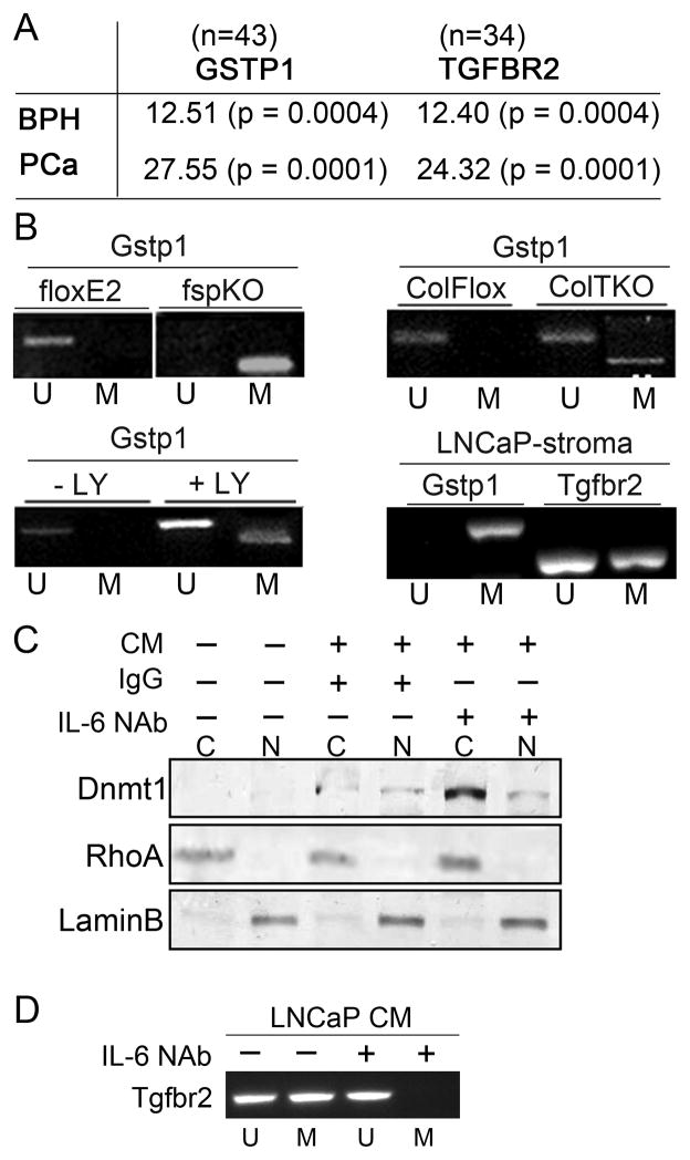
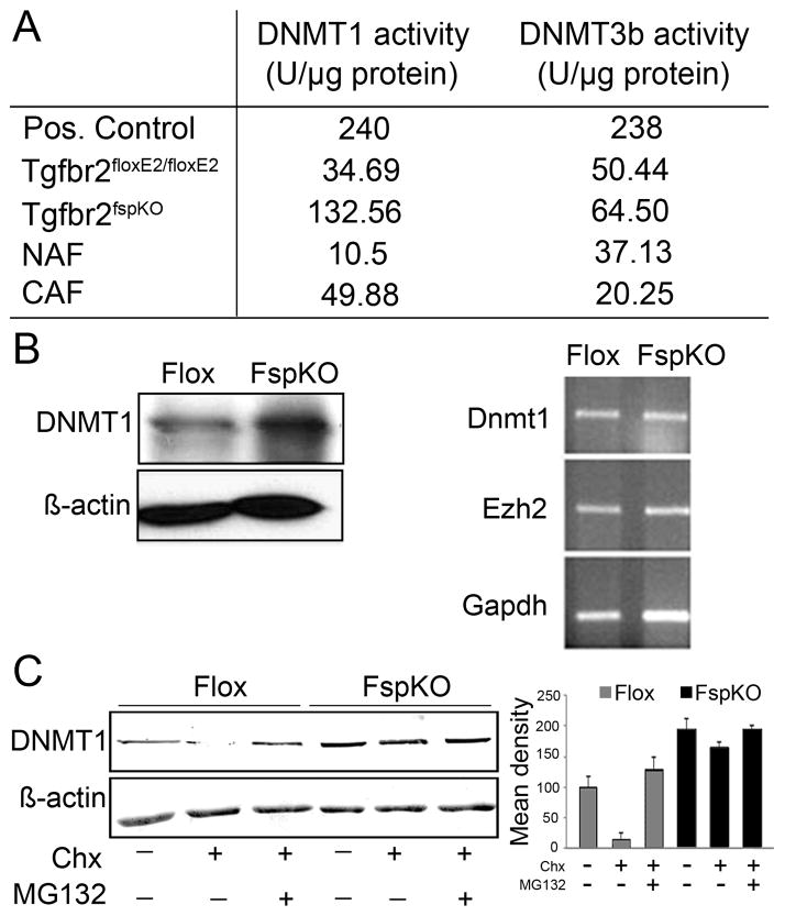
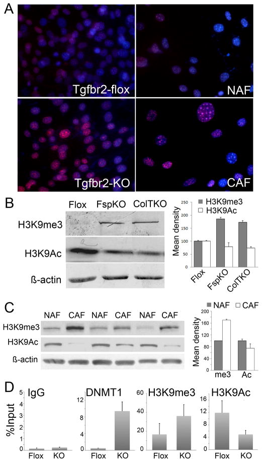
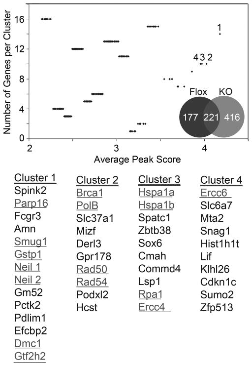
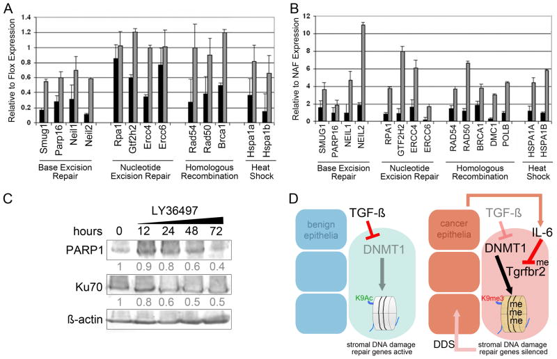
DNA damage found in prostate cancer-associated fibroblasts (CAF) promotes tumor progression. In the absence of somatic mutations in CAF, epigenetic changes dictate how stromal coevolution is mediated in tumors. Seventy percent of prostate cancer patients lose expression of transforming growth factor-beta type II receptor (TGFBR2) in the stromal compartment (n=77, P-value=0.0001), similar to the rate of glutathione S-transferase P1 (GSTP1) silencing. Xenografting of human prostate cancer epithelia, LNCaP, resulted in the epigenetic Tgfbr2 silencing of host mouse prostatic fibroblasts. Stromal Tgfbr2 promoter hypermethylation, initiated by LNCaP cells, was found to be dependent on interleukin 6 expression, based on neutralizing antibody studies. We further found that pharmacologic and transgenic knockout of TGF-β responsiveness in prostatic fibroblasts induced Gstp1 promoter methylation. It is known that TGF-β promotes DNA stability, however, the mechanism is not well understood. Both prostatic human CAF and mouse transgenic knockout of Tgbr2 had elevated DNA methyltransferase I (DNMT1) activity and histone H3 lysine 9 trimethylation (H3K9me3) to suggest greater promoter methylation. Interestingly, the conditional knockout of Tgfbr2 in mouse prostatic fibroblasts, in modeling epigenetic silencing of Tgfbr2, had greater epigenetic gene silencing of multiple DNA damage repair and oxidative stress response genes, based on promoter methylation array analysis. Homologous gene silencing was validated by reverse transcriptase (RT)-PCR in mouse and human prostatic CAF. Not surprisingly, DNA damage repair gene silencing in the prostatic stromal cells corresponded with the presence of DNA damage. Restoring the expression of the epigenetically silenced genes in wild-type fibroblasts with radiation-induced DNA damage reduced tumor progression. Tumor progression was inhibited even when epigenetic silencing was reversed in the Tgfbr2-knockout prostatic fibroblasts. Taken together, fibroblastic epigenetic changes causative of DNA damage, initiated by association with cancer epithelia, is a dominant mediator of tumor progression over TGF-β responsiveness.


Conflict of interest statement

Conflict of interest

The authors declare no conflicts of interest.

Figures

Figure 1
Figure 1. Prostate cancer mediate epigenetic changes in the associated stromal cells in a TGF-β-dependent manner
A. Promoter methylation analysis in BPH and PCa patient tissues support significant association between GSTP1 and TGFBR2 by Chi-square values and student T-test, in parenthesis. B. Gstp1 promoter methylation status in the prostatic fibroblastic cells from Tgfbr2floxE2/floxE2 and Tgfbr2fspKO mice were tested, as were Tgfbr2ColTKO prostatic stromal cells 72 hrs. following 4-OH tamoxifen-induced Cre activation by mPCR. Control Tgfbr2floxE2/floxE2 fibroblasts treated with or without the TGF-β reptor type I inhibitor, LY364947 (24h), mediated Gstp1 promoter methylation. Microdissected host CAF from LNCaP xenografts were tested for mouse Gstp1 and Tgfbr2 promoter methylation. Unmethylated (U) and methylated (M) DNA is indicated. C. Conditioned media (CM) from LNCaP cells was incubated with Tgfbr2floxE2/floxE2 fibroblasts for 2 days in the presence or absence of IgG and IL-6 neutralizing antibody (NAb). Cytoplasmic (C) and nuclear (N) fractions were Western blotted for Dnmt1 expression. RhoA and LaminB expression was used as loading controls for cytoplasmic and nuclear fractions, respectively. D. Tgbr2 promoter methylation status of prostatic fibroblasts by mPCR following LNCaP conditioned media incubation in the presence or absence of IL-6 NAb. The mPCR data in this figure is representative of six independent experiments with a significant densitometric difference between the treatment and control groups (p < 0.0001).
Figure 2
Figure 2. Post-translational regulation of DNMT1 expression
A. DNMT1 and DNMT3b activity was tested in Tgfbr2floxE2/floxE2 and Tgfbr2fspKO mouse prostatic stromal cells as well as human CAF and NAF cells. B. Western blotting indicated elevated DNMT1 protein expression in Tgfbr2fspKO, compared to Tgfbr2floxE2/floxE2 prostatic fibroblastic cells. However, RNA expression of DNMT1 and EZH2 were similarly expressed in Tgfbr2floxE2/floxE2 and Tgfbr2fspKO prostatic fibroblastic cells. C. Cyclohexamide (Chx) treatment resulted in the down regulation of DNMT1 protein expression in Tgfbr2floxE2/floxE2 prostatic fibroblasts. Antagonizing proteosome activity (MG132) restored DNMT1 expression in Chx treated cells. DNMT1 protein expression was unaffected by Chx or MG-132 in Tgfbr2fspKO prostatic fibroblasts. Columns in the graph are mean and SD of six densitometric readings adjusted for actin in independent blots prepared from three randomly-selected samples per treatment group (p < 0.0001).
Figure 3
Figure 3. Stromal histone and chromatin modification
A. H3K9me3 was immuno-localized in cultured mouse Tgfbr2-flox and Tgfbr2-KO prostatic fibroblasts as well as human NAF and CAF (red). Nuclei were counter stained with Hoechst (blue). B. H3K9me3 and H3K9Ac3 expression in prostatic fibroblastic cells from Tgfbr2floxE2/floxE2 and Tgfbr2fspKO mice, as well as those from Tgfbr2ColTKO mice 72 hours following tamoxifen-induced Cre activation were compared to β-actin loading control by Western blotting. Semi-quantitative densitometry of the bands from three independent Western blots showed significant differences in Tgfbr2-KO and Tgfbr2-Flox fibroblasts (p<0.0001). C. Protein expression of H3K9me3 and H2K9Ac in NAF and CAF differed significantly (p<0.001). Columns in the graph are mean values and standard deviation of six independent samples of NAF and CAF (only 3 of each are shown) adjusted for actin expression. D. DNMT1, H3K9me3, and H3K9Ac loading on the Gstp1 promoter were measured by ChIP analysis in Tgfbr2-flox and Tgfbr2-KO prostatic stromal cells by qPCR, relative to input DNA. Nonspecific IgG was used as a negative control.
Figure 4
Figure 4. Promoter methylation in prostatic fibroblast occur in a TGF-β-dependent manner
Methylation array revealed differences in Tgfbr2-KO (grey) and Tgfbr2-flox (black) prostatic fibroblasts. The Venn diagram illustrates the distribution of methylated genes in the two fibroblastic cell types validated by two independent array platforms. Cluster analysis indicated differential methylation of DNA-damage repair genes (underlined text, statistical significance set at p-value ≤ 0.05), with a summary of the specific gene names corresponding to the numbered clusters having the greatest peak scores.
Figure 5
Figure 5. Prostatic mouse Tgfbr2-KO and human CAF silencing of DNA damage repair genes are reversible by 5-aza-dC treatment
RNA expression of DNA damage repair genes were measured by qPCR. A. The expression in Tgfbr2-KO as compared to Tgfbr2-flox mouse fibroblastic cells following vehicle (black bars) and 5-aza-dC treatment (grey bar) was used to determine if promoter methylation affected gene silencing. All genes shown were significantly regulated by DNA methylation (p < 0.01), except Rpa1 and Ercc6. B. The expression of human homologs of the mouse genes were tested in CAF, as compared to NAF, following treatment with vehicle or 5-aza-dC. All genes shown were significantly regulated by DNA methylation (p < 0.01). C. Antagonizing TGF-β by LY36497 treatment of human NAF indicate regulation of PARP1 and Ku70 expression in a time course of 0 – 72 by Western blotting. The densitometry of the blots indicate relative PARP1 and Ku70 expression normalized to β-actin. D. A summary of our understanding of the mechanism by which stromal co-evolution contributes to a vicious cycle of stromal-epithelial interaction in cancer progression. The down regulated pathways are dimmed and active signaling are highlighted.

References

    1. Bhowmick NA, Chytil A, Plieth D, Gorska AE, Dumont N, Shappell S, et al. TGF-beta signaling in fibroblasts modulates the oncogenic potential of adjacent epithelia. Science. 2004;303(5659):848–51. Epub 2004/02/07. - PubMed
    1. Bhowmick NA, Neilson EG, Moses HL. Stromal fibroblasts in cancer initiation and progression. Nature. 2004;432(7015):332–7. Epub 2004/11/19. - PMC - PubMed
    1. Nguyen DX, Bos PD, Massague J. Metastasis: from dissemination to organ-specific colonization. Nat Rev Cancer. 2009;9(4):274–84. Epub 2009/03/25. - PubMed
    1. Tuxhorn JA, Ayala GE, Smith MJ, Smith VC, Dang TD, Rowley DR. Reactive stroma in human prostate cancer: induction of myofibroblast phenotype and extracellular matrix remodeling. Clin Cancer Res. 2002;8(9):2912–23. - PubMed
    1. Olumi AF, Grossfeld GD, Hayward SW, Carroll PR, Tlsty TD, Cunha GR. Carcinoma-associated fibroblasts direct tumor progression of initiated human prostatic epithelium. Cancer Res. 1999;59(19):5002–11. - PMC - PubMed

Publication types

MeSH terms

Substances