. 2013 Nov 13;32(22):2963-79.
doi: 10.1038/emboj.2013.222. Epub 2013 Oct 18.

A small RNA activates CFA synthase by isoform-specific mRNA stabilization

Affiliations

A small RNA activates CFA synthase by isoform-specific mRNA stabilization

Kathrin Sophie Fröhlich et al. EMBO J. .

Abstract

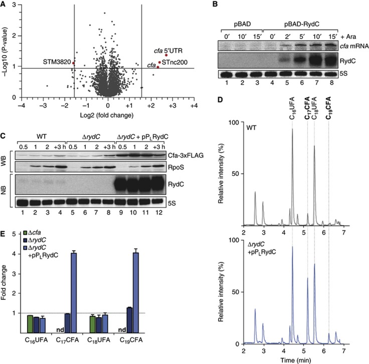
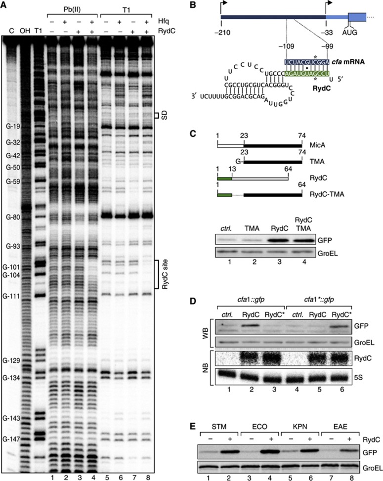
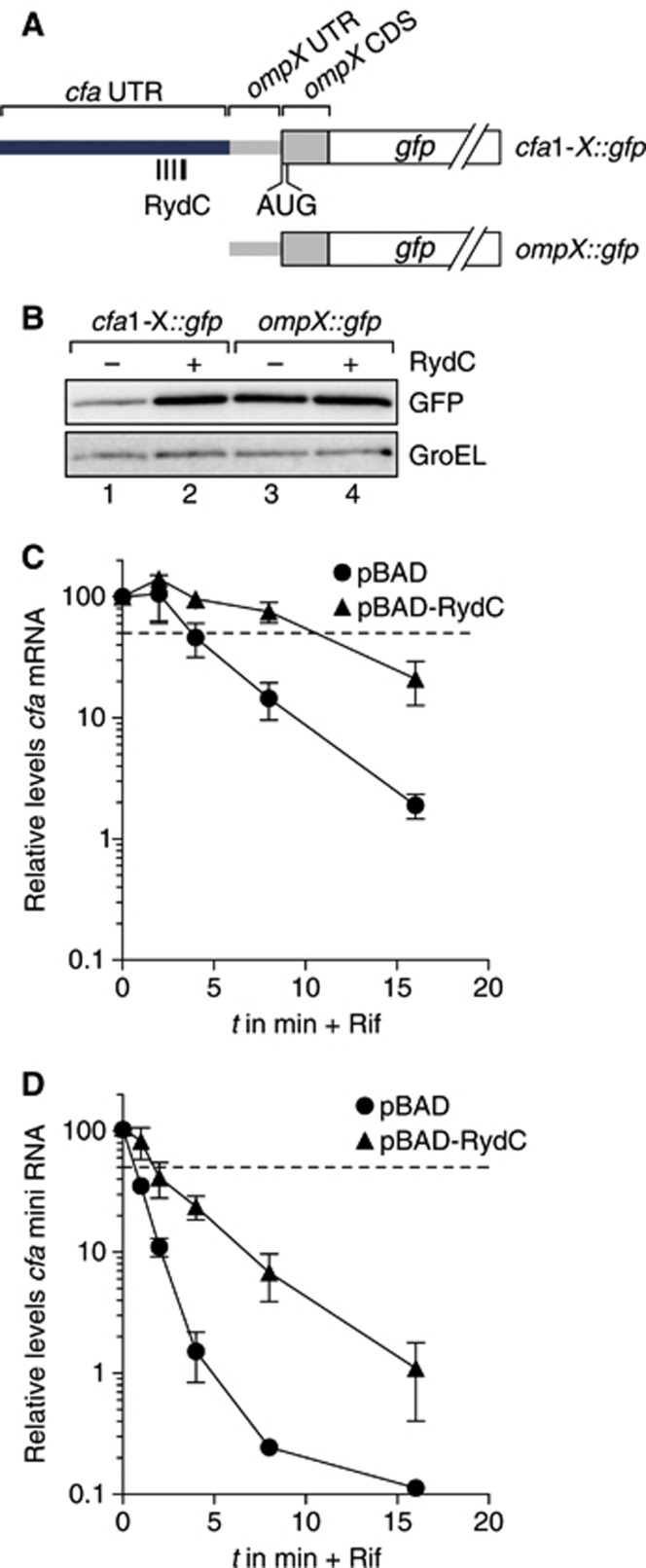
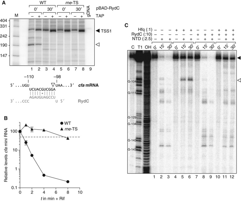
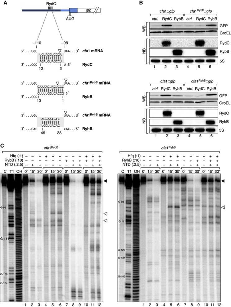
Small RNAs use a diversity of well-characterized mechanisms to repress mRNAs, but how they activate gene expression at the mRNA level remains not well understood. The predominant activation mechanism of Hfq-associated small RNAs has been translational control whereby base pairing with the target prevents the formation of an intrinsic inhibitory structure in the mRNA and promotes translation initiation. Here, we report a translation-independent mechanism whereby the small RNA RydC selectively activates the longer of two isoforms of cfa mRNA (encoding cyclopropane fatty acid synthase) in Salmonella enterica. Target activation is achieved through seed pairing of the pseudoknot-exposed, conserved 5' end of RydC to an upstream region of the cfa mRNA. The seed pairing stabilizes the messenger, likely by interfering directly with RNase E-mediated decay in the 5' untranslated region. Intriguingly, this mechanism is generic such that the activation is equally achieved by seed pairing of unrelated small RNAs, suggesting that this mechanism may be utilized in the design of RNA-controlled synthetic circuits. Physiologically, RydC is the first small RNA known to regulate membrane stability.


Conflict of interest statement

The authors declare that they have no conflict of interest.

Figures

Figure 1
Figure 1
Conservation, structure and expression of RydC. (A) Alignment and pseudoknot structure of RydC RNA. STM: Salmonella Typhimurium; SBG: Salmonella bongori; CKO: Citrobacter koseri; ECO: Escherichia coli; SFL: Shigella flexneri; EFE: Escherichia fergusoni; EAE: Enterobacter aerogenes; KPN: Klebsiella pneumoniae. Mutations K1, K2 and K1/2 introduced to alter pseudoknot formation are indicated. (B) RydC levels in total RNA samples (corresponding to 1 OD600) of wild-type Salmonella at indicated time points of growth were compared on northern blots to signals of in vitro transcribed RydC to estimate the in vivo copy number. 5S RNA served as a loading control. (C) Expression levels of RydC, RydC-K1, RydC-K2 and RydC-K1/2 (as described in (A); expressed from the constitutive PL promoter) were determined in ΔrydC Salmonella by northern blot analysis of total RNA samples (OD600 of 1). (D) Stabilities of RydC, RydC-K1, RydC-K2 and RydC-K1/2 were determined in ΔrydC Salmonella by northern blot analysis of total RNA samples withdrawn prior to and at indicated time points after inhibition of transcription by rifampicin at an OD600 of 1. See Supplementary Figure S4 for quantification. Source data for this figure is available on the online supplementary information page.
Figure 2
Figure 2
RydC induces cfa expression and activity. (A) Microarray analysis of Salmonella genes affected by pulse overexpression of RydC. RydC expression was induced by addition of arabinose (final concentration: 0.2%) to rydC mutant cells carrying pBAD-RydC or control plasmid pBAD. Changes in transcript abundances were scored on Salmonella-specific microarrays; genes displaying >3-fold change (P-value<0.15) are marked in red. (B) RydC and cfa mRNA levels were determined on northern blots of total RNA extracted from rydC mutant cells carrying plasmids pBAD or pBAD-RydC at indicated time points prior to and after addition of arabinose (Ara). The oligo directed against the 5′ UTR of cfa specifically recognizes cfa1 mRNA. (C) Expression of Cfa-3xFLAG in wild-type and ΔrydC mutant Salmonella either carrying a control construct or a plasmid for the constitutive overexpression of RydC from the PL promoter was monitored over growth on western blots. (D) Total ion chromatograms of Salmonella wild-type cells carrying a control plasmid or a ΔrydC mutant transformed with the RydC overexpression plasmid pPLRydC. Cells were grown in M9 minimal medium to exponential phase (OD600 of 0.5), and after alkaline hydrolysis, total fatty acids were analysed by LC/MS. Peaks assigned to C16UFA, C17CFA, C18UFA and C19CFA are indicated. (E) Relative quantification of C16UFA, C17CFA, C18UFA and C19CFA in Salmonella Δcfa or ΔrydC carrying either a control plasmid or pPLRydC. All measurements were normalized to wild type; error bars represent the standard deviation calculated from three independent biological replicates; nd: not detected. Source data for this figure is available on the online supplementary information page.
Figure 3
Figure 3
RydC acts on one of the two isoforms of cfa mRNA. (A) Sequence of the Salmonella cfa upstream region. Transcription initiates from two start sites indicated by arrows at −210 bp (σ70-dependent; cfa1 mRNA) or −33 bp (σS-dependent; cfa2 mRNA) relative to the translational start site, respectively. Promoter elements are highlighted in grey, and the start codon is boxed. (B) RydC specifically acts on the longer cfa mRNA isoform. Salmonella cfa::3xFLAG ΔrydC cells or an isogenic ΔrpoS mutant were transformed with the pBAD control plasmid (−) or the pBAD-RydC overexpression plasmid (+); Salmonella Δcfa ΔrydC served as a negative control. All strains were grown to an OD600 of 2, and total RNA samples withdrawn prior to and 15′ after arabinose addition were used as templates for primer extension. Transcripts originating from either TSS1 or TSS2 were identified using gene-specific sequencing ladders. (C) Schematic representation of the cfa gene including the upstream promoter region. Translational cfa::gfp fusions (under control of the constitutive PLTetO-1 promoter) were constructed comprising the 5′ upstream region from the distal (cfa1::gfp) or the proximal start site (cfa2::gfp) plus the first 45 nucleotides of the cfa CDS. (D) Regulation of reporter fusions was monitored by western blot analysis. At an OD600 of 1, total protein samples were prepared from Salmonella ΔrydC ΔrpoS mutants carrying plasmids to express cfa2::gfp or cfa1::gfp in combination with a control plasmid (−) or pPLRydC (+). GroEL served as a loading control. Expression of RydC was validated on a northern blot. Source data for this figure is available on the online supplementary information page.
Figure 4
Figure 4
RydC employs its conserved, single-stranded 5′ end to base pair with cfa1 mRNA. (A) In vitro structure probing using 5′ end-labelled cfa mRNA (TSS1 to nt 70 of the CDS; 20 nM) with lead(II) acetate (lanes 1–4) and RNase T1 (lanes 5–8) in the presence and absence of Hfq (20 nM) and RydC (200 nM). RNase T1 and alkaline ladders of cfa mRNA were used to map cleaved fragments. Positions of G-residues are indicated relative to the translational start site. The RydC binding site and the Shine-Dalgarno (SD) region are marked. (B) Predicted duplex forming between RydC (nts 2–11) and cfa mRNA (nts −109 to −99 relative to the translational start site). Positions of single-nucleotide exchanges generating the compensatory mutants RydC* and cfa* mRNA are indicated. (C) Schematic representation of wild-type MicA and RydC as well as the derivative constructs. The first 13 nts of RydC were fused to the 3′ part of MicA (nts 23–74; TMA) to construct RydC-TMA. The 5′ end of RydC is required to interact with cfa mRNA. GFP levels were determined on western blots of total protein samples isolated from Salmonella ΔrydC ΔrpoS mutants carrying plasmids for cfa1::gfp and either a control or plasmids for PL-driven overexpression of TMA, RydC or RydC-TMA. (D) Validation of the RydC-cfa mRNA interaction. Salmonella ΔrydC ΔrpoS mutants carrying plasmids for cfa1::gfp and cfa1*::gfp in combination with a control plasmid or RydC overexpression plasmids pPLRydC and pPLRydC*. Expression of GFP-fusion proteins was monitored on western blots of total protein samples prepared from cells in exponential growth (OD600 of 1). Equal expression of RydC and RydC* was controlled by northern blot analysis. (E) The regulation of cfa by RydC is conserved. GFP expression was monitored by western blot analysis in Salmonella ΔrydC mutant cells carrying translational Salmonella (STM), E. coli (ECO), K. pneumoniae (KPN) or E. aerogenes (EAE) cfa1::gfp reporter fusions in combination with either a control (−) or a plasmid to overexpress RydC versions of the indicated species (+). Source data for this figure is available on the online supplementary information page.
Figure 5
Figure 5
RydC can stabilize cfa mRNA. (A) Schematic representation of gfp fusion plasmids encompassing indicated fragments of the proximal cfa 5′ UTR (TSS1 to −62) inserted upstream of the 33-nt long 5′ UTR and the first 10 amino acids of ompX. (B) Regulation of reporter fusions as described in (A) was monitored by western blot analysis of total protein samples prepared from Salmonella ΔrydC ΔrpoS mutants in the absence (−) and presence (+) of RydC. (C) Stability of cfa mRNA in the presence of RydC. Salmonella ΔrydC ΔrpoS cells carrying either plasmids pBAD or pBAD-RydC were grown to OD600 of 1.0 when L-arabinose was added to induce RydC expression. After 15 min of induction, cultures were treated with rifampicin, and RNA samples were prepared from cells prior to and at indicated time points post treatment. Abundance of cfa mRNA was determined by qRT–PCR analysis. The signal obtained at 0 min was set to 100%, and the percentage of mRNA remaining at each time point was plotted on the y axis versus time on the x axis. The time point at which 50% of cfa mRNA had been decayed (dashed line) was calculated to determine the half-life (t1/2). Error bars represent the standard deviation calculated from three independent biological replicates. (D) Stability of cfa mini RNA in the presence of RydC. The cfa mini RNA was constitutively expressed in Salmonella ΔrydC Δcfa cells carrying either plasmids pBAD or pBAD-RydC, and its decay was analysed as described in (B). Source data for this figure is available on the online supplementary information page.
Figure 6
Figure 6
Binding of RydC interferes with RNase E cleavage of cfa mRNA. (A) 5′ RACE experiments were performed to determine the 5′ ends of cfa mRNA fragments in the absence and presence of RydC. Salmonella ΔrydC ΔrpoS Ptet-cfa::3xFLAG rne-ctrl (WT) and its isogenic rne-ts strain (rne-TS) carrying plasmid pBAD-RydC were grown at 44°C for 30 min. RNA isolated from culture samples withdrawn prior to and 30 min after induction of RydC was tobacco acid pyrophosphatase (TAP)-treated (+) or mock-treated (−) and used to prepare cDNA. Cleavage of the 5′ triphosphate group that is characteristic of primary transcripts by TAP renders the RNA a preferred substrate in the adjacent step of RNA linker ligation. A TAP-dependent elongation product (black arrowhead) and a second, TAP-independent PCR fragment (open arrowhead) were extracted, cloned and sequenced. Salmonella genomic DNA (gDNA) served as a negative control. The 5′ end of the cfa fragment (white arrowhead) is located within the region base-pairing RydC. (B) Inactivation of RNase E stabilizes the cfa mini RNA. The cfa mini RNA was constitutively expressed in Salmonella ΔrydC Ptet-cfa::3xFLAG rne-ctrl (WT) and its isogenic rne-ts strain (rne-TS) at the non-permissive temperature of 44°C. Decay of cfa mini RNA upon rifampicin addition was analysed as described in Figure 5B. (C) Mapping of cfa mRNA cleavage sites in vitro. Time-course experiment monitoring RNase E-mediated decay of 5′ labelled in vitro transcribed cfa mRNA fragment (TSS1 to −72). In all, 20 nM of mRNA was incubated with 50 nM purified RNase E N-terminal catalytic domain (NTD) in the absence or presence of Hfq (20 nM), and RydC or RydC* sRNAs (200 nM) as indicated. Reactions were stopped prior to and 15 or 30 min post addition of NTD. RNase T1 and alkaline ladders of cfa mRNA were employed to map cleavage products. The cleavage intermediate of cfa mRNA is indicated by an open arrowhead and mapped to position −99 relative to the cfa translational start site. Source data for this figure is available on the online supplementary information page.
Figure 7
Figure 7
(A) Schematic representation of the base-pairing regions in cfa1, cfa1RybB or cfa1RyhB reporter fusions and the expected base-pairing interactions to RydC, RybB or RyhB, respectively. RNase E cleavage sites identified in in vitro assays are indicated by open arrowheads. (B) The regulation of cfa expression is independent of the actual seed sequence. Salmonella ΔrydC ΔrybB or ΔryhB mutants were transformed with cfa1::gfp and either cfa1RybB::gfp or cfa1RyhB::gfp reporter fusions, respectively. GFP levels were determined by western blot analysis in the presence of either a control plasmid or constructs to constitutively overexpress RydC, RybB or RyhB. See Supplementary Figure S9 for quantification. Expression of RydC, RybB and RyhB sRNAs was monitored on northern blots. (C) Determination of cfa1RybB (left panel) and cfa1RyhB (right panel) cleavage sites in vitro. Time-course experiment of RNase E-mediated decay of cfa1 variants as described in Figure 6C but using cfa1RybB or cfa1RyhB RNA and RybB or RyhB sRNAs, respectively. Mapping of the cleavage intermediates indicated by an open arrowhead is marked in (A). Source data for this figure is available on the online supplementary information page.
Figure 8
Figure 8
RydC interacts with one of the two isoforms of cfa mRNA to increase Cfa. Two independent promoter sites controlled by either σ70 or the alternative σ-factor σS control the transcription of cfa. By base pairing with its conserved 5′ end to the longer of the two mRNA isoforms, RydC increases cfa1 mRNA stability and levels. The increased protein levels lead to an alteration of membrane stability, as Cfa converts the double bond of unsaturated fatty acid side chains into cyclopropane rings.

References

    1. Afonyushkin T, Vecerek B, Moll I, Blasi U, Kaberdin VR (2005) Both RNase E and RNase III control the stability of sodB mRNA upon translational inhibition by the small regulatory RNA RyhB. Nucleic Acids Res 33: 1678–1689 - PMC - PubMed
    1. Antal M, Bordeau V, Douchin V, Felden B (2005) A small bacterial RNA regulates a putative ABC transporter. J Biol Chem 280: 7901–7908 - PubMed
    1. Argaman L, Hershberg R, Vogel J, Bejerano G, Wagner EG, Margalit H, Altuvia S (2001) Novel small RNA-encoding genes in the intergenic regions of Escherichia coli. Curr Biol 11: 941–950 - PubMed
    1. Arnold TE, Yu J, Belasco JG (1998) mRNA stabilization by the ompA 5′ untranslated region: two protective elements hinder distinct pathways for mRNA degradation. RNA 4: 319–330 - PMC - PubMed
    1. Bandyra KJ, Said N, Pfeiffer V, Gorna MW, Vogel J, Luisi BF (2012) The seed region of a small RNA drives the controlled destruction of the target mRNA by the endoribonuclease RNase E. Mol Cell 47: 943–953 - PMC - PubMed

Publication types