Review
. 2013 Nov;31(11):999-1008.
doi: 10.1038/nbt.2725. Epub 2013 Oct 20.

Reassessing target antigens for adoptive T-cell therapy

Affiliations
Review

Reassessing target antigens for adoptive T-cell therapy

Christian S Hinrichs et al. Nat Biotechnol. 2013 Nov.

Abstract

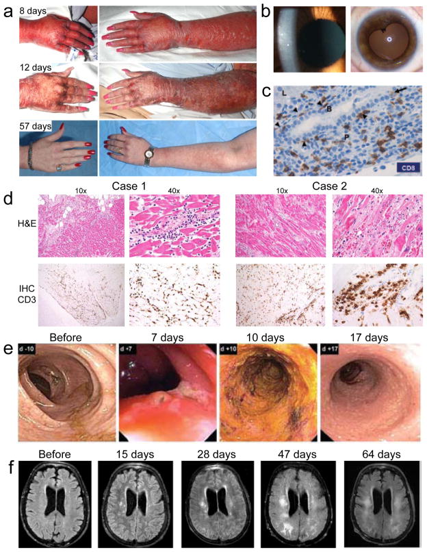
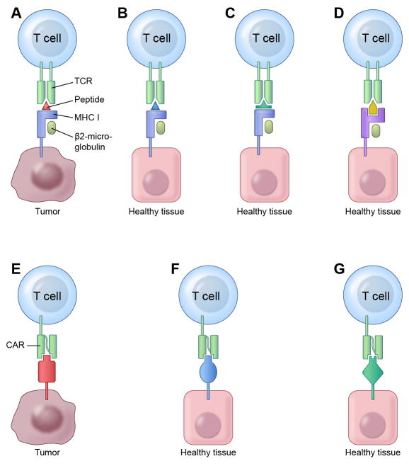
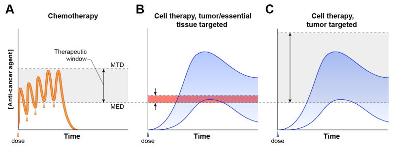
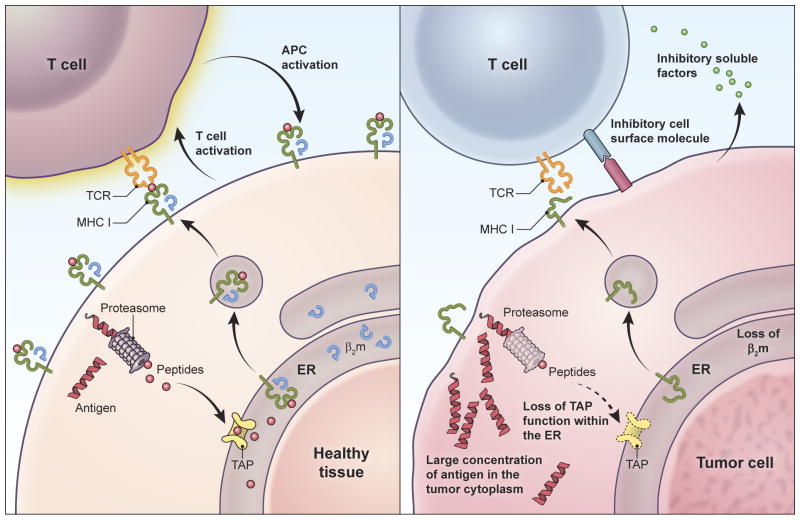
Adoptive T-cell therapy can target and kill widespread malignant cells thereby inducing durable clinical responses in melanoma and selected other malignances. However, many commonly targeted tumor antigens are also expressed by healthy tissues, and T cells do not distinguish between benign and malignant tissues if both express the target antigen. Autoimmune toxicity from T cell-mediated destruction of normal tissue has limited the development and adoption of this otherwise promising type of cancer therapy. A review of the unique biology of T-cell therapy and of recent clinical experience compels a reassessment of target antigens that traditionally have been viewed from the perspective of weaker immunotherapeutic modalities. It is important that target antigens chosen for adoptive T-cell therapy are expressed by tumors and not by essential healthy tissues. The risk of adverse autoimmune events can be further mitigated by generating antigen receptors using strategies that reduce the chance of cross-reactivity against epitopes in unintended targets. In general, a circumspect approach to target selection and thoughtful preclinical and clinical studies are pivotal to the ongoing advancement of these promising treatments.


Figures

Figure 1
Figure 1
Autoimmune adverse events in ACT clinical trials. (a) Skin rash at various time points after treatment of a melanoma patient with T cells engineered to express a TCR with high affinity for MART1. (b) Cellular anterior chamber infiltrate in a melanoma patient two weeks after treatment with T cells engineered to express a TCR with high affinity for MART1 (left). Aymptomatic posterior synechiae six months after treatment of the same patient (right). (c) Immunohistochemical (IHC) analysis of CD8 expression in a liver biopsy obtained four days after treatment of a renal cell carcinoma patient with T cells transduced with a CAR specific for carbonic anhydrase IX. CD8 T cells line the basal side of (arrowheads) and infiltrate (arrow) the bile duct epithelium. Liver parenchyma (L), portal triangle (P), and bile duct (B) are labeled. (d) Colonoscopy images from a colon cancer patient at various time points after administration of T cells engineered to express a TCR specific for carcinoembryonic antigen (CEA). Transient severe colitis is evident. (e) Magnetic resonance imaging of the brain of a melanoma patient after various time points after injection of T cells expressing a receptor that recognizes MAGE-A3 but is cross-reactive with MAGE-A12. Images show progressive white matter changes consistent with leukomalacia.
Figure 2
Figure 2
Scenarios in which TCRs and CARs can recognize and cross-react with unintended target antigens,. (a-d) TCRs recognize target antigen peptides bound to MHC molecules. (a) The ideal tumor antigen targeted by adoptively transferred T cells is a peptide-MHC complex that is expressed uniquely by tumor cells and is not expressed by healthy tissue. (b-f) However, even T cells engineered to target such ideal tumor antigens may recognize and damage healthy tissue in certain scenarios. (b) Healthy tissue may express distinct antigens that contain peptide epitopes identical to the one recognized by the engineered T cells. Vigilance for this type of cross-reactivity is particularly important when targeting peptides in antigens encoded by genes arising from gene duplication such as some CTA family members (e.g. MAGE). (c) Healthy tissue may express distinct antigens that contain peptide epitopes that are different in sequence than the intended target peptide, but sufficiently similar in structure to bind to the same MHC molecule and be recognized by the TCR on the engineered T cells. (d) Healthy tissue may express complexes of different MHC molecules with peptides from non-target antigens that are cross-recognized by the engineered TCR. (e-g) CARs directly engage intact target antigens, rather than antigen-derived peptides bound to MHC molecules. (e) An ideal CAR for ACT specifically recognizes a single epitope that is unique to a tumor-restricted antigen. (f-g) However, in some scenarios even T cells engineered to express such ideal CARs can recognize unintended target antigens. (f) Non-target antigens expressed by healthy tissues may contain epitopes identical to the intended CAR target and therefore be recognized by the CAR-expressing T cells. (g) Antibody specificity for an epitope can be imperfect, permitting structurally similar (but different in sequence) epitopes present on non-target antigens to be cross-recognized by a CAR targeting a tumor-specific antigen.
Figure 3
Figure 3
Differences in the pharmacokinetics and mechanisms of action between cytotoxic chemotherapy and adoptive T cell therapies have important implications for phase I clinical trials and determination of therapeutic range. (a) Patient-to-patient variation in drug levels (orange line) is small for chemotherapeutics. Drug exposure (the area under the curve) is predictable and, in the case of toxicity, can be terminated by stopping drug dosing (each dose is indicated by an arrowhead). The efficacy of chemotherapy derives from the greater sensitivity of tumors than vital normal tissues to the cytotoxic actions of the drug. This difference in tissue sensitivity creates a therapeutic window (gray shaded area) between the minimum effective dose (MED) (the dose at which tumor regression occurs) and the maximum tolerated dose (MTD) (the dose at which intolerable toxicities occur) of the drug. (b-c) Because engineered T cells used in ACT are produced on a patient-to-patient basis their pharmacokinetics fluctuate substantially (blue shaded area). Given this variability, it is not clear that the MTD from one cohort of patients can be broadly applied to others. Furthermore, safe phase I testing is challenging because infused T cells do not degrade predictably but rather – unlike any other type of cancer treatment – increase in quantity (due to T cell proliferation) and can persist indefinitely (due to formation of long-lived memory T cell populations) following infusion. Therefore, in the case of adverse events, exposure to T cell therapies cannot be terminated reliably by the standard approach of stopping drug dosing ,. (b) A therapeutic window (red shaded area) in which durable regression of high-tumor-burden disease occurs without debilitating autoimmunity has thus far not been identified for any cellular therapy directed against antigens expressed by tumors and essential healthy tissues–,,. (c) Targeting an antigen that is not expressed by healthy tissue opens the therapeutic window (gray shaded area, by increasing the MTD) for ACT because direct cytotoxicity to normal tissues does not occur, even at high doses of cells.
Figure 4
Figure 4
T cells may target healthy tissues more efficiently than tumors, independent of the relative abundance of target antigen on each tissue. This phenomenon may preclude identification of a therapeutic window based on enhanced sensitivity of tumor compared to healthy tissue to T cell-mediated killing. Because high-avidity T cells have exquisite sensitivity for their target antigens and can recognize even single complexes of target peptide-MHC, healthy tissues expressing even small quantities of antigen but possessing intact antigen processing and presentation machinery might be highly vulnerable to T cell-mediated killing (left panel). Once initiated, such T cell killing can be amplified by the proliferation of T cells and by the mutual activation of T cells and antigen presenting cells. Despite expressing potentially large quantities of target antigen, tumors are prone to genomic instability, defects in antigen processing and presentation (e.g. defects in transporter associated with antigen processing function (TAP), proteosomal subunits, β-2 microglobulin, and MHC molecules) (right panel). As such they may not present target antigen to engineered T cells. Tumors may be further protected by their production of cell surface and soluble molecules that inhibit T cell activation.

References

    1. Cheever MA, et al. The prioritization of cancer antigens: a National Cancer Institute pilot project for the acceleration of translational research. Clin Cancer Res. 2009;15:5323–5337. - PMC - PubMed
    1. Rosenberg SA, Yang JC, Restifo NP. Cancer immunotherapy: moving beyond current vaccines. Nat Med. 2004;10:909–915. - PMC - PubMed
    1. Rosenberg SA, et al. Durable complete responses in heavily pretreated patients with metastatic melanoma using T-cell transfer immunotherapy. Clin Cancer Res. 2011;17:4550–4557. - PMC - PubMed
    1. Robbins PF, et al. Tumor regression in patients with metastatic synovial cell sarcoma and melanoma using genetically engineered lymphocytes reactive with NY-ESO-1. J Clin Oncol. 2011;29:917–924. - PMC - PubMed
    1. Kochenderfer JN, et al. B-cell depletion and remissions of malignancy along with cytokine-associated toxicity in a clinical trial of anti-CD19 chimeric-antigen-receptor-transduced T cells. Blood. 2012;119:2709–2720. - PMC - PubMed

MeSH terms