. 2013 Dec;195(24):5639-50.
doi: 10.1128/JB.00990-13. Epub 2013 Oct 18.

ExsA and LcrF recognize similar consensus binding sites, but differences in their oligomeric state influence interactions with promoter DNA

Affiliations

ExsA and LcrF recognize similar consensus binding sites, but differences in their oligomeric state influence interactions with promoter DNA

Jessica M King et al. J Bacteriol. 2013 Dec.

Abstract

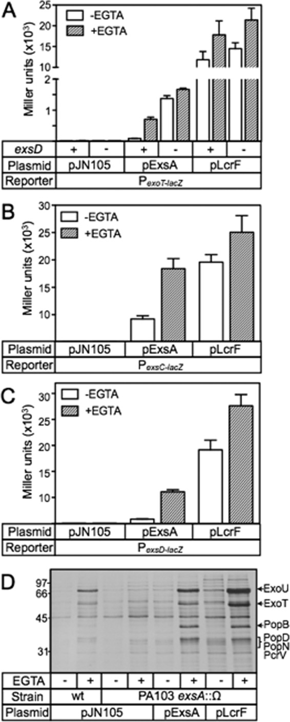
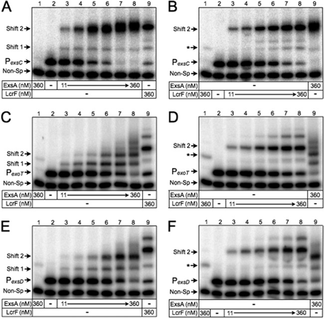
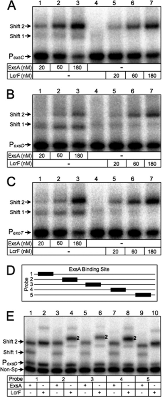
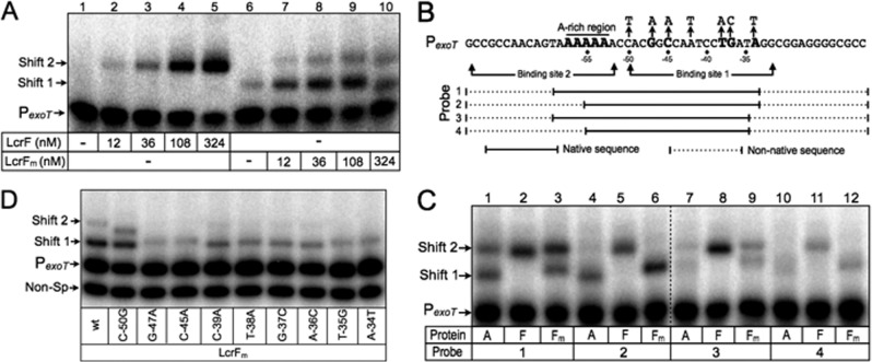
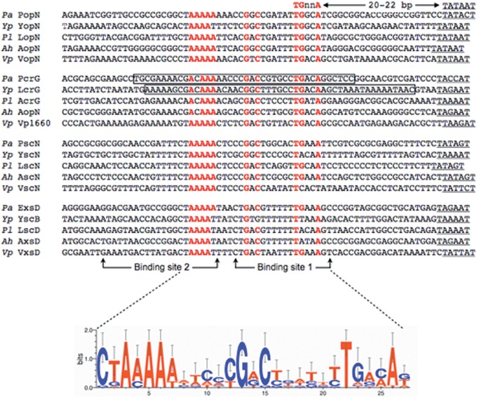
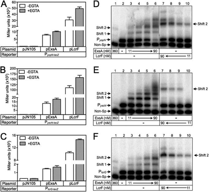
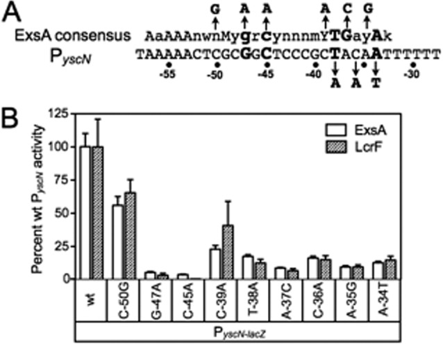
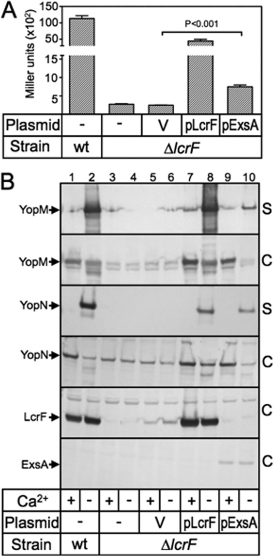
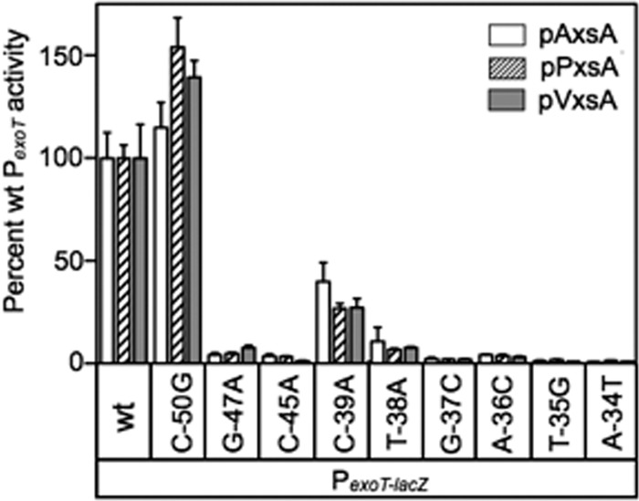
ExsA activates type III secretion system (T3SS) gene expression in Pseudomonas aeruginosa and is a member of the AraC family of transcriptional regulators. AraC proteins contain two helix-turn-helix (HTH) DNA binding motifs. One helix from each HTH motif inserts into the major groove of the DNA to make base-specific contacts with the promoter region. The amino acids that comprise the HTH motifs of ExsA are nearly identical to those in LcrF/VirF, the activators of T3SS gene expression in the pathogenic yersiniae. In this study, we tested the hypothesis that ExsA/LcrF/VirF recognize a common nucleotide sequence. We report that Yersinia pestis LcrF binds to and activates transcription of ExsA-dependent promoters in P. aeruginosa and that plasmid-expressed ExsA complements a Y. pestis lcrF mutant for T3SS gene expression. Mutations that disrupt the ExsA consensus binding sites in both P. aeruginosa and Y. pestis T3SS promoters prevent activation by ExsA and LcrF. Our combined data demonstrate that ExsA and LcrF recognize a common nucleotide sequence. Nevertheless, the DNA binding properties of ExsA and LcrF are distinct. Whereas two ExsA monomers are sequentially recruited to the promoter region, LcrF binds to promoter DNA as a preformed dimer and has a higher capacity to bend DNA. An LcrF mutant defective for dimerization bound promoter DNA with properties similar to ExsA. Finally, we demonstrate that the activators of T3SS gene expression from Photorhabdus luminescens, Aeromonas hydrophila, and Vibrio parahaemolyticus are also sensitive to mutations that disrupt the ExsA consensus binding site.


Figures

Fig 1
Fig 1
LcrF complements an exsA mutant for T3SS gene expression. (A to C) The PA103 exsA::Ω strain carrying either the PexoT-lacZ (A), PexsC-lacZ (B), or PexsD-lacZ (C) transcriptional reporter was transformed with a vector control (pJN105), an ExsA expression vector (pExsA), or an LcrF expression vector (pLcrF). The resulting strains were cultured under noninducing (−EGTA, open bars) or inducing (+EGTA, hatched bars) conditions for T3SS gene expression and assayed for β-galactosidase activity as reported in Miller units. *, P < 0.001; ** P < 0.01. (D) Silver-stained gel of concentrated culture supernatant fluid prepared from wild-type PA103 or the PA103 exsA::Ω strain carrying the indicated plasmids following growth under noninducing (−EGTA) or inducing (+EGTA) conditions for T3SS gene expression. The positions of molecular mass standards are indicated on the left side of the gel, and the type III secreted proteins ExoU, ExoT, PopB, PopD, PopN, and PcrV are labeled on the right side of the gel (49).
Fig 2
Fig 2
DNA binding properties of purified ExsA and LcrF. EMSAs were performed using radiolabeled probes derived from the ExsA-dependent PexsC (A and B), PexoT (C and D), and PexsD (E and F) promoters. A nonspecific probe (Non-Sp) derived from the algD promoter region was included in all binding reactions as a negative control. Probes (0.05 nM each) were incubated in the absence or presence of 11, 23, 45, 90, 180, or 360 nM ExsA (A, C, and E) or LcrF (B, D, and F) for 15 min at 25°C. Samples were analyzed by native polyacrylamide gel electrophoresis and phosphorimaging. The positions of shift products 1 and 2 are indicated. The asterisk indicates background shifting of the nonspecific probe by LcrF.
Fig 3
Fig 3
DNA bending properties of ExsA and LcrF. (A to C) EMSAs using 50-bp radiolabeled probes derived from the ExsA-dependent PexsC (A), PexsD (B), and PexoT (C) promoters. Probes (0.05 nM each) were incubated in the presence of 20, 60, or 180 nM ExsA (lanes 1 to 3 in each panel) or LcrF (lanes 5 to 7 in each panel) for 15 min at 25°C. Samples were analyzed by native polyacrylamide gel electrophoresis and phosphorimaging. (D) Diagram depicting the position of the ExsA binding site (black box) derived from the PexsD promoter within probes 1 to 5 (solid line). (E) Circular permutation experiment performed using probes 1 to 5 (0.05 nM each) incubated in the presence of 180 nM ExsA (odd-numbered lanes) or LcrF (even-numbered lanes) for 15 min at 25°C. Samples were analyzed by native polyacrylamide gel electrophoresis and phosphorimaging. The positions of shift products 1 and 2 are indicated.
Fig 4
Fig 4
ExsA- and LcrF-dependent activation is sensitive to nucleotide substitutions in the ExsA consensus site. (A) Sequence of the PexoT promoter showing the conserved GnC and TGnnA sequences (highlighted in bold) and the nucleotide substitutions indicated with an arrow. (B) The PA103 exsA::Ω strain carrying the indicated PexoT-lacZ reporters was transformed with pExsA or pLcrF. The resulting strains were cultured in the presence of EGTA and assayed for β-galactosidase activity. Activation by ExsA (open bars) and LcrF (hatched bars) is reported as the percent activity normalized to the activity of the wild-type PexoT-lacZ reporter. (C and D) EMSAs using radiolabeled probes derived from the mutant PexoT promoters. The nonspecific PalgD probe (Non-Sp) was included as a negative control. Probes (0.05 nM each) were incubated in the presence of 45 nM ExsA (C) or LcrF (D) for 15 min at 25°C. Samples were analyzed by native polyacrylamide gel electrophoresis and phosphorimaging. The positions of shift products 1 and 2 are indicated.
Fig 5
Fig 5
LcrF binds to the P. aeruginosa PexoT promoter probe as a dimer. (A) EMSA using 50-bp radiolabeled probes derived from the ExsA-dependent PexoT promoter. Probes (0.05 nM each) were incubated in the presence of 12, 36, 108, 324 nM LcrF (lanes 2 to 5) or LcrFm (lanes 6 to 10) for 15 min at 25°C. Samples were analyzed by native polyacrylamide gel electrophoresis and phosphorimaging. (B) Diagram depicting PexoT promoter probes with truncations that destroy binding sites 1 and/or 2. Solid and dotted lines represent native and nonnative PexoT sequences, respectively. The adenine-rich region and conserved GnC and TGnnA sequences are indicated in bold. (C) EMSA using 60-bp radiolabeled probes derived from the ExsA-dependent PexoT promoter. Probes (0.05 nM each) were incubated in the presence of 90 nM ExsA(A), LcrF(F), or LcrFm for 15 min at 25°C. Samples were analyzed and imaged as described above.
Fig 6
Fig 6
Sequence alignment of the ExsA consensus binding sequence in ExsA (Pseudomonas aeruginosa)-, LcrF (Yersinia pestis)-, PxsA (Photorhabdus luminescens)-, AxsA (Aeromonas hydrophila)-, and VxsA (Vibrio parahaemolyticus)-dependent promoters. ExsA binding sites 1 and 2 are indicated with arrows. The consensus sequence is indicated in red, and −10 regions are underlined. The boxed regions in the P. aeruginosa PpcrG and Y. pestis PlcrG promoters correspond to regions protected by ExsA and LcrF/VirF from DNase I cleavage, respectively (see Fig. S5 in the supplemental material) (31, 48).
Fig 7
Fig 7
ExsA activates transcription of Y. pestis transcriptional reporters. (A to C) The PA103 exsA::Ω strain carrying either the PyopN-lacZ (A), PyscN-lacZ (B), or PlcrG-lacZ (C) reporters was transformed with pJN105, pExsA, or pLcrF. The resulting strains were cultured under noninducing (−EGTA, open bars) or inducing (+EGTA, hatched bars) conditions for T3SS gene expression and assayed for β-galactosidase activity. Values were reported in Miller units. (D to F) EMSAs using radiolabeled probes derived from the LcrF-dependent PyopN (D), PyscN (E), and PlcrG (F) promoters. The PalgD probe was included in all binding reactions as a nonspecific (Non-Sp) control. Probes (0.05 nM each) were incubated in the absence (lane 2) or presence of 11, 23, 45, or 90 nM ExsA (lanes 3 to 6 in each panel) or LcrF (lanes 7 to 10 in each panel) for 15 min at 25°C. Samples were analyzed by native polyacrylamide gel electrophoresis and phosphorimaging. The positions of shift products 1 and 2 are indicated.
Fig 8
Fig 8
Y. pestis PyscN reporter activity is sensitive to substitutions that disrupt the ExsA binding site. (A) The PyscN promoter sequence showing the ExsA consensus sequence in binding site 1. The GnC and TGnnA sequences are highlighted in bold, and each promoter nucleotide substitution is indicated with an arrow. (B) The PA103 exsA::Ω strain carrying the indicated mutant PyscN-lacZ transcriptional reporters was transformed with either pExsA or pLcrF. The resulting strains were cultured in the presence of EGTA and assayed for β-galactosidase activity. Activation by pExsA (open bars) and pLcrF (hatched bars) is reported as the percent activity of the mutant promoters normalized to the activity at the wild-type PyscN promoter.
Fig 9
Fig 9
ExsA activates expression of the Y. pestis T3SS. (A) Y. pestis KIM5-3001 (yopM::lacZYA) and KIM5-3233-F2 (ΔlcrF yopM::lacZYA) carrying a vector control (V), pLcrF, or pExsA were cultured under inducing conditions for T3SS gene expression and then assayed for β-galactosidase activity as reported in Miller units. (B) Wild-type Y. pestis KIM5-3001 (lanes 1 and 2) or KIM5-3001-F1 (ΔlcrF) (lanes 3 to 10) lacking a vector (lanes 1 to 4) or carrying a vector control (lanes 5 and 6), pLcrF (lanes 7 and 8), or pExsA (lanes 9 and10) were grown under noninducing conditions (+calcium) or inducing (−calcium; odd-numbered lanes) conditions for T3SS gene expression. Concentrated supernatant fluid (S) and cell-associated fractions (C) were prepared and subjected to SDS-PAGE and immunoblot analysis. Arrows indicate protein products.
Fig 10
Fig 10
The PA103 exsA::Ω strain carrying the indicated PexoT-lacZ transcriptional reporters was transformed with AxsA (pAxsA), PxsA (pPxsA), or VxsA (pVxsA) expression vectors. The resulting strains were cultured in the presence of EGTA and assayed for β-galactosidase activity. Activation by pAxsA (open bars), pPxsA (hatched bars), and pVxsA (gray bars) is reported as the percent activity of the mutant promoters normalized to the activity of the wild-type PexoT promoter.

Similar articles

Cited by

References

    1. Hauser AR. 2009. The type III secretion system of Pseudomonas aeruginosa: infection by injection. Nat. Rev Microbiol. 7:654–665 - PMC - PubMed
    1. Buttner D. 2012. Protein export according to schedule: architecture, assembly, and regulation of type III secretion systems from plant- and animal-pathogenic bacteria. Microbiol. Mol. Biol. Rev. 76:262–310 - PMC - PubMed
    1. Matsumoto H, Young GM. 2009. Translocated effectors of Yersinia. Curr. Opin. Microbiol. 12:94–100 - PMC - PubMed
    1. Engel J, Balachandran P. 2009. Role of Pseudomonas aeruginosa type III effectors in disease. Curr. Opin. Microbiol. 12:61–66 - PubMed
    1. Hayes CS, Aoki SK, Low DA. 2010. Bacterial contact-dependent delivery systems. Annu. Rev. Genet. 44:71–90 - PubMed

Publication types

MeSH terms

LinkOut - more resources