

A new addition to the cell plan of anammox bacteria: "Candidatus Kuenenia stuttgartiensis" has a protein surface layer as the outermost layer of the cell
- PMID: 24142254
- PMCID: PMC3911120
- DOI: 10.1128/JB.00988-13
A new addition to the cell plan of anammox bacteria: "Candidatus Kuenenia stuttgartiensis" has a protein surface layer as the outermost layer of the cell
Abstract
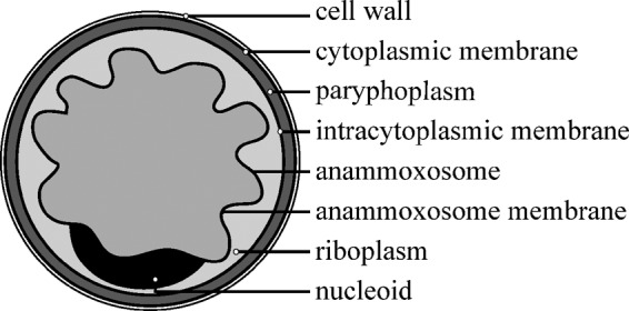
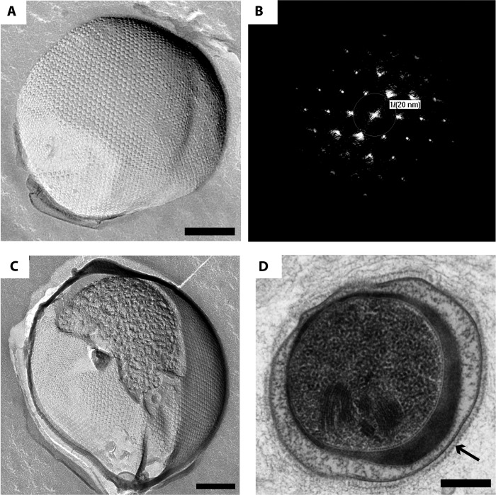
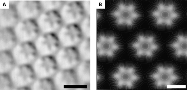
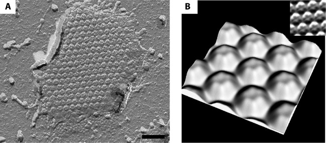
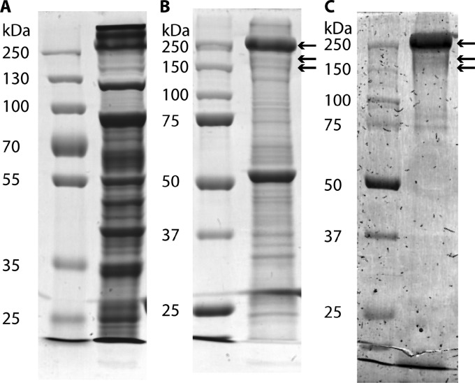
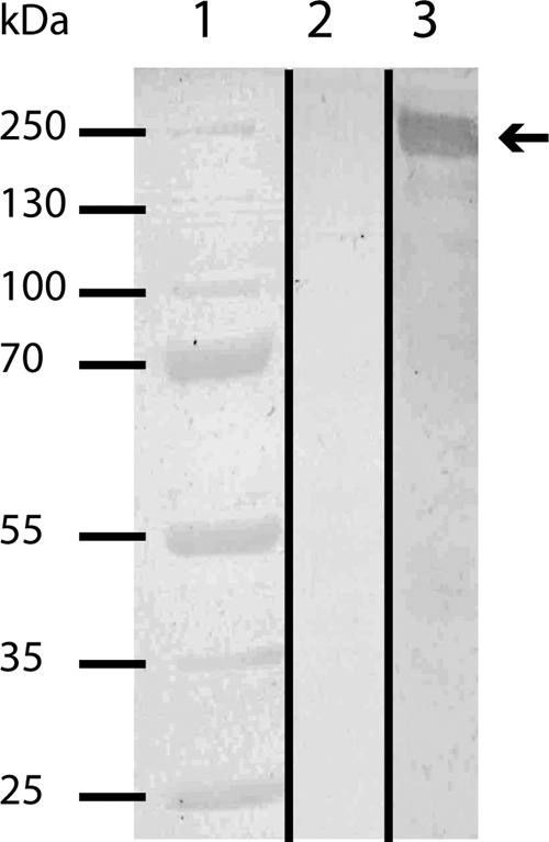
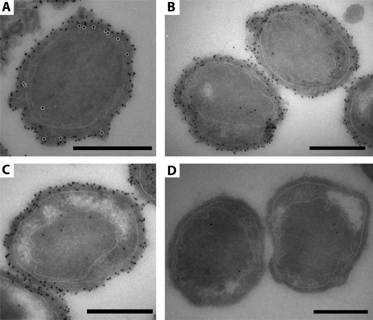
Anammox bacteria perform anaerobic ammonium oxidation (anammox) and have a unique compartmentalized cell consisting of three membrane-bound compartments (from inside outwards): the anammoxosome, riboplasm, and paryphoplasm. The cell envelope of anammox bacteria has been proposed to deviate from typical bacterial cell envelopes by lacking both peptidoglycan and a typical outer membrane. However, the composition of the anammox cell envelope is presently unknown. Here, we investigated the outermost layer of the anammox cell and identified a proteinaceous surface layer (S-layer) (a crystalline array of protein subunits) as the outermost component of the cell envelope of the anammox bacterium "Candidatus Kuenenia stuttgartiensis." This is the first description of an S-layer in the phylum of the Planctomycetes and a new addition to the cell plan of anammox bacteria. This S-layer showed hexagonal symmetry with a unit cell consisting of six protein subunits. The enrichment of the S-layer from the cell led to a 160-kDa candidate protein, Kustd1514, which has no homology to any known protein. This protein is present in a glycosylated form. Antibodies were generated against the glycoprotein and used for immunogold localization. The antiserum localized Kustd1514 to the S-layer and thus verified that this protein forms the "Ca. Kuenenia stuttgartiensis" S-layer.
Figures
Similar articles
-
Intracellular localization of membrane-bound ATPases in the compartmentalized anammox bacterium 'Candidatus Kuenenia stuttgartiensis'.Mol Microbiol. 2010 Aug;77(3):701-15. doi: 10.1111/j.1365-2958.2010.07242.x. Epub 2010 Jun 9. Mol Microbiol. 2010. PMID: 20545867 Free PMC article.
-
Characterization of the first planctomycetal outer membrane protein identifies a channel in the outer membrane of the anammox bacterium Kuenenia stuttgartiensis.Biochim Biophys Acta Biomembr. 2018 Mar;1860(3):767-776. doi: 10.1016/j.bbamem.2017.12.020. Epub 2017 Dec 27. Biochim Biophys Acta Biomembr. 2018. PMID: 29288627
-
The S-Layer Protein of the Anammox Bacterium Kuenenia stuttgartiensis Is Heavily O-Glycosylated.Front Microbiol. 2016 Nov 1;7:1721. doi: 10.3389/fmicb.2016.01721. eCollection 2016. Front Microbiol. 2016. PMID: 27847504 Free PMC article.
-
Cell biology of unique anammox bacteria that contain an energy conserving prokaryotic organelle.Antonie Van Leeuwenhoek. 2013 Oct;104(4):489-97. doi: 10.1007/s10482-013-9990-5. Epub 2013 Aug 9. Antonie Van Leeuwenhoek. 2013. PMID: 23929088 Review.
-
Anaerobic ammonium-oxidizing bacteria: unique microorganisms with exceptional properties.Microbiol Mol Biol Rev. 2012 Sep;76(3):585-96. doi: 10.1128/MMBR.05025-11. Microbiol Mol Biol Rev. 2012. PMID: 22933561 Free PMC article. Review.
Cited by
-
A Comparison of Anammox Bacterial Abundance and Community Structures in Three Different Emerged Plants-Related Sediments.Curr Microbiol. 2015 Sep;71(3):421-7. doi: 10.1007/s00284-015-0851-5. Epub 2015 Jun 16. Curr Microbiol. 2015. PMID: 26077223
-
Identification and characterization of an abundant lipoprotein from Methylacidiphilum fumariolicum SolV.Arch Microbiol. 2023 Jun 12;205(7):261. doi: 10.1007/s00203-023-03603-y. Arch Microbiol. 2023. PMID: 37306788 Free PMC article.
-
Surface-layer protein is a public-good matrix exopolymer for microbial community organisation in environmental anammox biofilms.ISME J. 2023 Jun;17(6):803-812. doi: 10.1038/s41396-023-01388-y. Epub 2023 Mar 4. ISME J. 2023. PMID: 36871068 Free PMC article.
-
Kolteria novifilia, a novel planctomycetotal strain from the volcanic habitat of Panarea divides by unusual lateral budding.J Bacteriol. 2025 Jul 24;207(7):e0033724. doi: 10.1128/jb.00337-24. Epub 2025 Jun 24. J Bacteriol. 2025. PMID: 40552805 Free PMC article.
-
Identification and characterisation of a major outer membrane protein from Methylacidiphilum fumariolicum SolV.Antonie Van Leeuwenhoek. 2023 Nov;116(11):1227-1245. doi: 10.1007/s10482-023-01879-0. Epub 2023 Sep 22. Antonie Van Leeuwenhoek. 2023. PMID: 37737555 Free PMC article.
References
Publication types
MeSH terms
Substances
LinkOut - more resources
Full Text Sources
Other Literature Sources
