

The potential role of genetically-modified pig mesenchymal stromal cells in xenotransplantation
- PMID: 24142483
- PMCID: PMC3946698
- DOI: 10.1007/s12015-013-9478-8
The potential role of genetically-modified pig mesenchymal stromal cells in xenotransplantation
Abstract
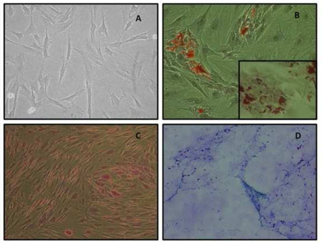
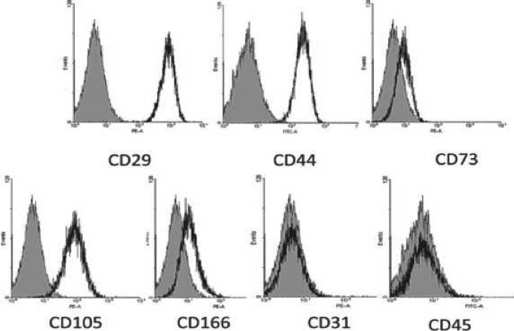
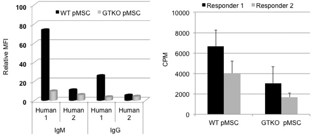
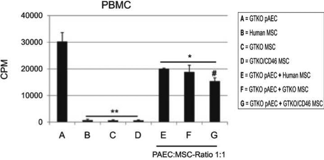
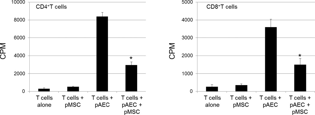
Mesenchymal stromal cells (MSCs) are known to have regenerative, anti-inflammatory, and immunodulatory effects. There are extensive indications that pig MSCs function satisfactorily across species barriers. Pig MSCs might have considerable therapeutic potential, particularly in xenotransplantation, where they have several potential advantages. (i) pMSCs can be obtained from the specific organ- or cell-source donor pig or from an identical (cloned) pig. (ii) They are easy to obtain in large numbers, negating the need for prolonged ex vivo expansion. (iii) They can be obtained from genetically-engineered pigs, and the genetic modification can be related to the therapeutic goal of the MSCs. We have reviewed our own studies on MSCs from genetically-engineered pigs, and summarize them here. We have successfully harvested and cultured MSCs from wild-type and genetically-engineered pig bone marrow and adipose tissue. We have identified several pig (p)MSC surface markers (positive for CD29, CD44, CD73, CD105, CD166, and negative for CD31, CD45), have demonstrated their proliferation and differentiation (into adipocytes, osteoblasts, and chondroblasts), and evaluated their antigenicity and immune suppressive effects on human peripheral blood mononuclear cells and CD4(+)T cells. They have identical or very similar characteristics to MSCs from other mammals. Genetically-modified pMSCs are significantly less immunogenic than wild-type pMSCs, and downregulate the human T cell response to pig antigens as efficiently as do human MSCs. We hypothesized that pMSCs can immunomodulate human T cells through induction of apoptosis or anergy, or cause T cell phenotype switching with induction of regulatory T cells, but we could find no evidence for these mechanisms. However, pMSCs upregulated the expression of CD69 on human CD4(+) and CD8(+) T cells, the relevance of which is currently under investigation. We conclude that MSCs from genetically-engineered pigs should continue to be investigated for their immunomodulatory (and regenerative and anti-inflammatory) effects in pig-to-nonhuman primate organ and cell transplantation models.
Conflict of interest statement
DA is an employee of Revivicor Inc., Blacksburg, VA. The other authors report no conflicts of interest.
Figures
Similar articles
-
Human T cells upregulate CD69 after coculture with xenogeneic genetically-modified pig mesenchymal stromal cells.Cell Immunol. 2013 Sep-Oct;285(1-2):23-30. doi: 10.1016/j.cellimm.2013.08.004. Epub 2013 Aug 29. Cell Immunol. 2013. PMID: 24044963 Free PMC article.
-
Genetically-modified pig mesenchymal stromal cells: xenoantigenicity and effect on human T-cell xenoresponses.Xenotransplantation. 2011 May-Jun;18(3):183-95. doi: 10.1111/j.1399-3089.2011.00635.x. Xenotransplantation. 2011. PMID: 21696448
-
Adipose-derived mesenchymal stromal cells from genetically modified pigs: immunogenicity and immune modulatory properties.Cytotherapy. 2012 Apr;14(4):494-504. doi: 10.3109/14653249.2011.651529. Epub 2012 Jan 23. Cytotherapy. 2012. PMID: 22264190 Free PMC article.
-
Do mesenchymal stem cells function across species barriers? Relevance for xenotransplantation.Xenotransplantation. 2012 Sep-Oct;19(5):273-85. doi: 10.1111/xen.12000. Xenotransplantation. 2012. PMID: 22978461 Free PMC article. Review.
-
Mesenchymal stem cells: Identification, phenotypic characterization, biological properties and potential for regenerative medicine through biomaterial micro-engineering of their niche.Methods. 2016 Apr 15;99:62-8. doi: 10.1016/j.ymeth.2015.09.016. Epub 2015 Sep 15. Methods. 2016. PMID: 26384580 Review.
Cited by
-
Translational Animal Models Provide Insight Into Mesenchymal Stromal Cell (MSC) Secretome Therapy.Front Cell Dev Biol. 2021 Mar 19;9:654885. doi: 10.3389/fcell.2021.654885. eCollection 2021. Front Cell Dev Biol. 2021. PMID: 33869217 Free PMC article. Review.
-
EP4 Antagonist-Elicited Extracellular Vesicles from Mesenchymal Stem Cells Rescue Cognition/Learning Deficiencies by Restoring Brain Cellular Functions.Stem Cells Transl Med. 2019 Jul;8(7):707-723. doi: 10.1002/sctm.18-0284. Epub 2019 Mar 19. Stem Cells Transl Med. 2019. PMID: 30891948 Free PMC article.
-
Xenogeneic and Stem Cell-Based Therapy for Cardiovascular Diseases: Genetic Engineering of Porcine Cells and Their Applications in Heart Regeneration.Int J Mol Sci. 2020 Dec 18;21(24):9686. doi: 10.3390/ijms21249686. Int J Mol Sci. 2020. PMID: 33353186 Free PMC article. Review.
-
Cardiac Xenotransplantation: Challenges, Evolution, and Advances.JACC Basic Transl Sci. 2022 Jun 15;7(7):716-729. doi: 10.1016/j.jacbts.2022.05.003. eCollection 2022 Jul. JACC Basic Transl Sci. 2022. PMID: 35958689 Free PMC article. Review.
-
Functional augmentation of naturally-derived materials for tissue regeneration.Ann Biomed Eng. 2015 Mar;43(3):555-67. doi: 10.1007/s10439-014-1192-4. Epub 2014 Nov 25. Ann Biomed Eng. 2015. PMID: 25422160 Free PMC article. Review.
References
-
- Friedenstein AJ, Petrakova KV, Kurolesova AI, Frolova GP. Heterotopic of bone marrow. Analysis of precursor cells for osteogenic and hematopoietic tissues. Transplantation. 1968;6:230–247. - PubMed
-
- Sordi V. Mesenchymal stem cell homing capacity. Transplantation. 2009;87:S42–S45. - PubMed
-
- Dominici M, Le Blanc K, Mueller I, Slaper-Cortenbach I, Marini F, Krause D, et al. Minimal criteria for defining multipotent mesenchymal stromal cells. The International Society for Cellular Therapy position statement. Cytotherapy. 2006;8:315–317. - PubMed
Publication types
MeSH terms
Substances
Grants and funding
LinkOut - more resources
Full Text Sources
Other Literature Sources
Molecular Biology Databases
Research Materials
Miscellaneous
