Review
. 2013 Dec;305(12):L912-33.
doi: 10.1152/ajplung.00259.2013. Epub 2013 Oct 18.

Airway smooth muscle in airway reactivity and remodeling: what have we learned?

Affiliations
Review

Airway smooth muscle in airway reactivity and remodeling: what have we learned?

Y S Prakash. Am J Physiol Lung Cell Mol Physiol. 2013 Dec.

Abstract

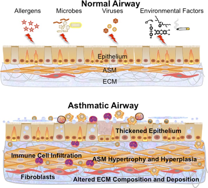
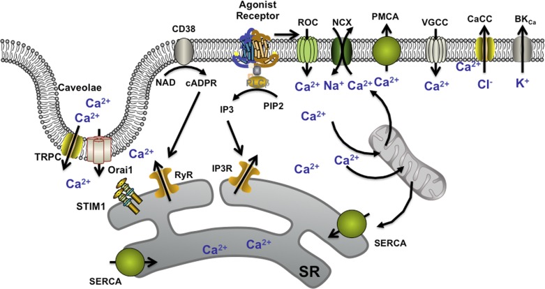
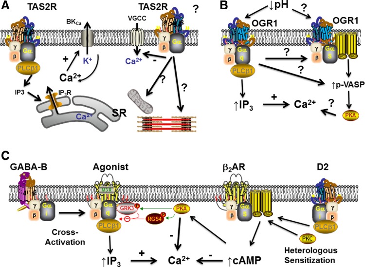
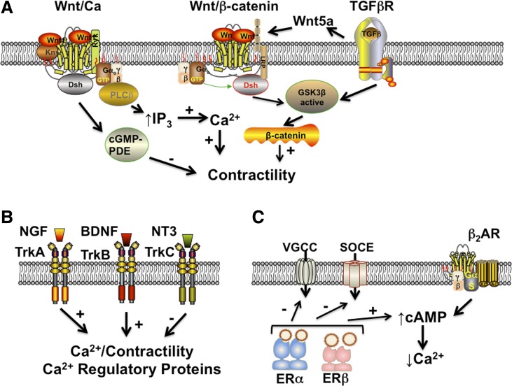
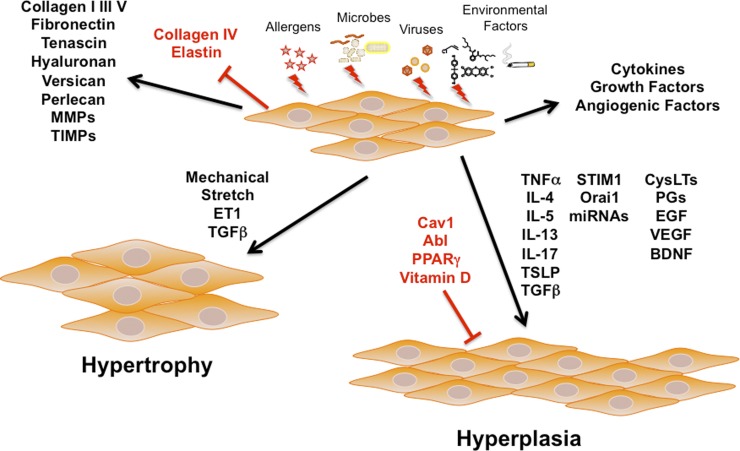
It is now established that airway smooth muscle (ASM) has roles in determining airway structure and function, well beyond that as the major contractile element. Indeed, changes in ASM function are central to the manifestation of allergic, inflammatory, and fibrotic airway diseases in both children and adults, as well as to airway responses to local and environmental exposures. Emerging evidence points to novel signaling mechanisms within ASM cells of different species that serve to control diverse features, including 1) [Ca(2+)]i contractility and relaxation, 2) cell proliferation and apoptosis, 3) production and modulation of extracellular components, and 4) release of pro- vs. anti-inflammatory mediators and factors that regulate immunity as well as the function of other airway cell types, such as epithelium, fibroblasts, and nerves. These diverse effects of ASM "activity" result in modulation of bronchoconstriction vs. bronchodilation relevant to airway hyperresponsiveness, airway thickening, and fibrosis that influence compliance. This perspective highlights recent discoveries that reveal the central role of ASM in this regard and helps set the stage for future research toward understanding the pathways regulating ASM and, in turn, the influence of ASM on airway structure and function. Such exploration is key to development of novel therapeutic strategies that influence the pathophysiology of diseases such as asthma, chronic obstructive pulmonary disease, and pulmonary fibrosis.

Keywords: asthma; bronchoconstriction; bronchodilation; calcium; development; extracellular matrix; inflammation; lung; proliferation.


Figures

Fig. 1.
Fig. 1.
Transformations toward the asthmatic airway. Exposure of the normal airway to insults such as allergens, microbes, or viruses or to environmental factors such as pollutants, tobacco smoke, or nanoparticles results in changes throughout the epithelium, airway smooth muscle (ASM), and extracellular matrix (ECM). The asthmatic airway involves infiltration of a variety of immune cells, a thickened epithelium with goblet cell hyperplasia, increased mucus, a thickened, more fibrotic ASM layer with increased cell size (hypertrophy) and numbers (hyperplasia), along with altered ECM composition. Changes within the ASM layer can be a result of processes initiated or modulated by as well as involving ASM cells.
Fig. 2.
Fig. 2.
Mechanisms of [Ca2+]i regulation in ASM. A number of regulatory mechanisms are well recognized, including agonist-induced G-protein coupled receptor (GPCR)-based production of inositol trisphosphate (IP3) and action of the ectoenzyme CD38 in producing the second messenger cyclic ADP ribose (cADPR), leading to sarcoplasmic reticulum (SR) Ca2+ release from IP3 receptor and ryanodine receptor (RyR) channels, respectively. Additionally, Ca2+ influx can occur through receptor-operated channels (ROC), voltage-gated Ca2+ channels (VGCCs), and perhaps through the bidirectional Na+/Ca2+ exchanger (NCX). Depletion of SR Ca2+ stores can lead to store-operated Ca2+ entry (SOCE) mediated by the SR Ca2+-sensing protein stromal interacting molecule (STIM1) and the plasma membrane influx channels Orai1 and canonical transient receptor potential (TRPC) (both being expressed with caveolae). Elevated [Ca2+]i can be sequestered by the SR via the Ca2+ ATPase (SERCA), by mitochondrial buffering, and plasma membrane efflux mechanisms such as the plasma membrane Ca2+ ATPase (PMCA) or by membrane hyperpolarization due to BKCa and Ca2+-activated chloride channels (CaCC). PLC, phospholipase C; cADPR, cyclic ADP ribose; PIP2, phosphatidylinositol 4,5-bisphosphate.
Fig. 3.
Fig. 3.
Novel GPCR-based signaling mechanisms in ASM. A: effect of bitter tastants such as chloroquine and saccharin on bitter taste receptor (TAS2R) GPCRs in ASM has been a topic of substantial recent interest. Working through a Gq protein-coupled mechanism (where the α-subunit may represent gustducin, transducin, or α1), TAS2R activation leads to increased SR Ca2+ release via IP3 receptor channels and elevated [Ca2+]i levels but intriguingly results in bronchodilation via mechanisms that remain to be established. Ca2+-activated K+ channels (BKCa) and inhibition of voltage-gated Ca2+ channels are thought to be involved, but pathways such as inhibition of other Ca2+ influx mechanisms, mitochondria, or contractile mechanisms downstream to Ca2+ may also be relevant. B: a novel, intriguing GPCR mechanism recently identified in ASM is an extracellular pH-sensitive pathway, ovarian cancer G protein-coupled receptor 1 (OGR1/GPR68), that on the one hand appears to work via Gq to increase [Ca2+]i via the IP3 mechanism, but on the other hand also activates the protein kinase A cascade either directly through Gq or perhaps through interactions with Gs. VASP, vasodilator-stimulated phosphoprotein. C: in contrast to the novel TAS2R and OGR1 pathways, there is now increasing evidence that the GABA and dopaminergic systems (well known in neuroscience) also have interesting effects in ASM. Activation of the metabotropic GABA-B receptor results in a Gi-coupled cross activation of Gq-coupled receptors of bronchoconstrictor agonists, leading to increased [Ca2+]i. On the other hand, prolonged dopaminergic signaling via a D2-like, Gi-coupled pathway leads to enhancement of the Gs-coupled β2-adrenoceptor (β2AR) system, thus increasing cAMP and promoting bronchodilation. GRK3, G protein-coupled receptor kinase; RGS4, G-protein signaling 4.
Fig. 4.
Fig. 4.
Novel signaling cascades in ASM. A: activation of transforming growth factor (TGF)-β receptors in ASM leads to increased expression of Wnt5a that can in turn activate either the Wnt/GSK3β/β-catenin system in promoting contractility or alternatively the noncanonical Wnt/Ca system that increases [Ca2+]i via IP3. Conversely, the Wnt/Ca system could modulate contractility by activating cGMP-specific phosphodiesterases (PDE). B: although well recognized in the nervous system, there is now considerable evidence that the receptor tyrosine kinase family of neurotrophic receptors (tropomyosin-related kinases; Trks) are expressed and functional in ASM. The neurotrophin nerve growth factor (NGF) acts via TrkA, whereas brain-derived neurotrophic factor (BDNF) acts through TrkB to enhance [Ca2+]i and expression of Ca2+ regulatory proteins to promote contractility. Conversely, neurotrophin-3 (NT3) acts via TrkC to blunt Ca2+ and contractility. C: given sex differences in allergic airway diseases, the mechanisms by which sex steroids such as estrogens can influence ASM are being explored. Limited data suggest that estrogen receptors (ERs) can inhibit VGCC and SOCE to reduce [Ca2+]i while separately increasing cAMP that also aids in bronchodilation, perhaps by potentiating β2AR effects.
Fig. 5.
Fig. 5.
Role of ASM in remodeling. Besides its role in modulating contractility, there is now substantial evidence that insults such as allergens, infection, and environmental factors can modulate the “synthetic” aspects of ASM. Here, ASM can produce a range of ECM proteins including collagen, fibronectin, the matrix metalloproteinases (MMPs), and their tissue inhibitors (TIMPs), as well as a number of pro- and anti-inflammatory cytokines, growth factors (e.g., BDNF), and angiogenic factors (e.g., vascular endothelial growth factor, VEGF). Furthermore, in response to insults, autocrine/paracrine effects of locally produced factors (e.g., cytokines and growth factors) and intracellular mechanisms (e.g., STIM1, Orai1, miRNAs), ASM cells can increase in size (hypertrophy) or number (hyperplasia), thus contributing to increased ASM mass noted in asthma. PPAR, peroxisome proliferator-activated receptor; TSLP, thymic stromal lymphopoietin; ET1, endothelin 1; PGs, prostaglandins.

References

    1. Algara-Suarez P, Mejia-Elizondo R, Sims SM, Saavedra-Alanis VM, Espinosa-Tanguma R. The 13 isoform of Na+-Ca2+ exchanger expressed in guinea pig tracheal smooth muscle is less sensitive to KB-R7943. J Physiol Biochem 66: 117–125, 2010 - PubMed
    1. Algara-Suarez P, Romero-Mendez C, Chrones T, Sanchez-Armass S, Meza U, Sims SM, Espinosa-Tanguma R. Functional coupling between the Na+/Ca2+ exchanger and nonselective cation channels during histamine stimulation in guinea pig tracheal smooth muscle. Am J Physiol Lung Cell Mol Physiol 293: L191–L198, 2007 - PubMed
    1. Alrashdan YA, Alkhouri H, Chen E, Lalor DJ, Poniris M, Henness S, Brightling CE, Burgess JK, Armour CL, Ammit AJ, Hughes JM. Asthmatic airway smooth muscle CXCL10 production: mitogen-activated protein kinase JNK involvement. Am J Physiol Lung Cell Mol Physiol 302: L1118–L1127, 2012 - PMC - PubMed
    1. An SS, Wang WC, Koziol-White CJ, Ahn K, Lee DY, Kurten RC, Panettieri RA, Jr, Liggett SB. TAS2R activation promotes airway smooth muscle relaxation despite β(2)-adrenergic receptor tachyphylaxis. Am J Physiol Lung Cell Mol Physiol 303: L304–L311, 2012 - PMC - PubMed
    1. Aravamudan B, Thompson M, Pabelick C, Prakash YS. Brain-derived neurotrophic factor induces proliferation of human airway smooth muscle cells. J Cell Mol Med 16: 812–823, 2012 - PMC - PubMed

Publication types

MeSH terms

LinkOut - more resources