

Kch1 family proteins mediate essential responses to endoplasmic reticulum stresses in the yeasts Saccharomyces cerevisiae and Candida albicans
- PMID: 24142703
- PMCID: PMC3843098
- DOI: 10.1074/jbc.M113.508705
Kch1 family proteins mediate essential responses to endoplasmic reticulum stresses in the yeasts Saccharomyces cerevisiae and Candida albicans
Abstract
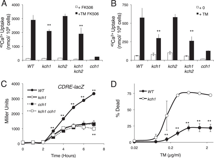
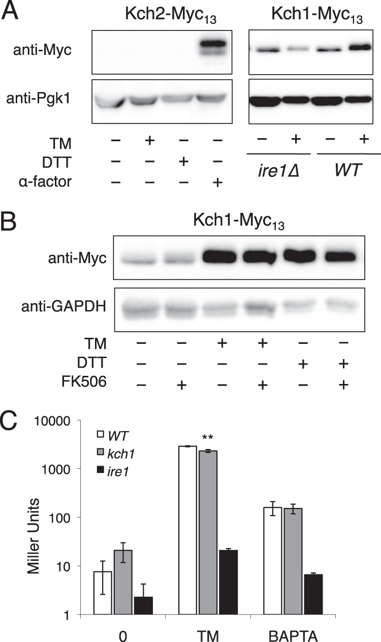
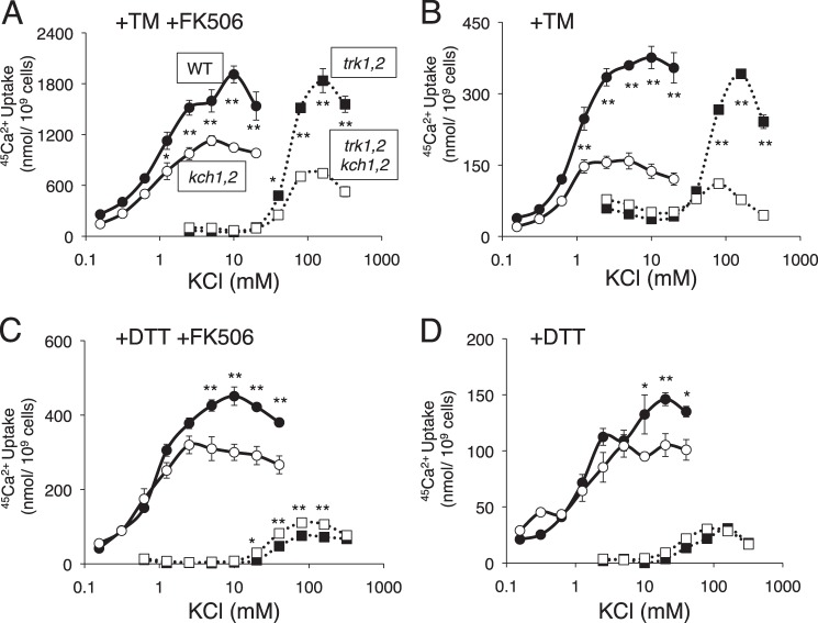
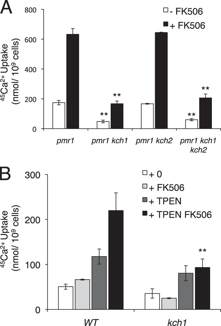
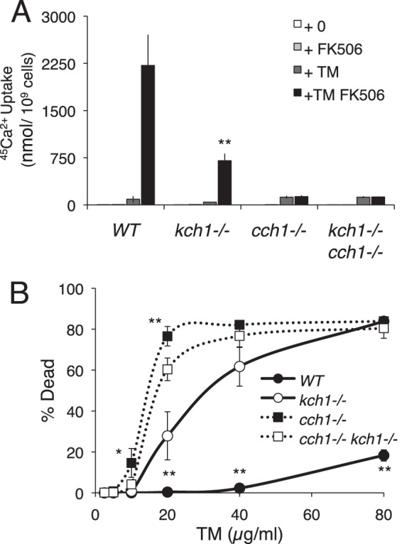
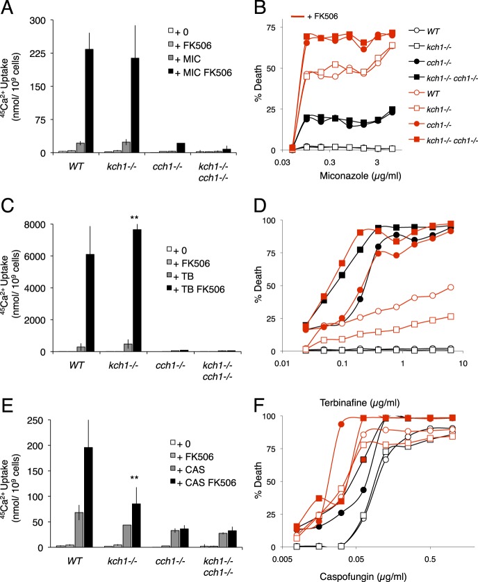
The activation of a high affinity Ca(2+) influx system (HACS) in the plasma membrane is required for survival of yeast cells exposed to natural or synthetic inhibitors of essential processes (secretory protein folding or sterol biosynthesis) in the endoplasmic reticulum (ER). The mechanisms linking ER stress to HACS activation are not known. Here we show that Kch1, a recently identified low affinity K(+) transporter in the plasma membrane of Saccharomyces cerevisiae, is up-regulated in response to several ER stressors and necessary for HACS activation. The activation of HACS required extracellular K(+) and was also dependent on the high affinity K(+) transporters Trk1 and Trk2. However, a paralog of Kch1 termed Kch2 was not expressed and not necessary for HACS activation in these conditions. The pathogenic yeast Candida albicans carries only one homolog of Kch1/Kch2, and homozygous knock-out mutants were similarly deficient in the activation of HACS during the responses to tunicamycin. However, the Kch1 homolog was not necessary for HACS activation or cell survival in response to several clinical antifungals (azoles, allylamines, echinocandins) that target the ER or cell wall. Thus, Kch1 family proteins represent a conserved linkage between HACS and only certain classes of ER stress in these yeasts.
Keywords: Antifungals; Calcineurin; Calcium Channels; Cell Death; ER Stress; Potassium Transport.
Figures
Similar articles
-
Activation of an essential calcium signaling pathway in Saccharomyces cerevisiae by Kch1 and Kch2, putative low-affinity potassium transporters.Eukaryot Cell. 2013 Feb;12(2):204-14. doi: 10.1128/EC.00299-12. Epub 2012 Nov 30. Eukaryot Cell. 2013. PMID: 23204190 Free PMC article.
-
Yeast Kch1 and Kch2 membrane proteins play a pleiotropic role in membrane potential establishment and monovalent cation homeostasis regulation.FEMS Yeast Res. 2017 Aug 1;17(5). doi: 10.1093/femsyr/fox053. FEMS Yeast Res. 2017. PMID: 28810704
-
New regulators of a high affinity Ca2+ influx system revealed through a genome-wide screen in yeast.J Biol Chem. 2011 Mar 25;286(12):10744-54. doi: 10.1074/jbc.M110.177451. Epub 2011 Jan 20. J Biol Chem. 2011. PMID: 21252230 Free PMC article.
-
Monovalent cation transporters at the plasma membrane in yeasts.Yeast. 2019 Apr;36(4):177-193. doi: 10.1002/yea.3355. Epub 2018 Oct 3. Yeast. 2019. PMID: 30193006 Review.
-
The coupling of plasma membrane calcium entry to calcium uptake by endoplasmic reticulum and mitochondria.J Physiol. 2014 Jan 15;592(2):261-8. doi: 10.1113/jphysiol.2013.255661. Epub 2013 Jun 24. J Physiol. 2014. PMID: 23798493 Free PMC article. Review.
Cited by
-
Identification of Genes in Candida glabrata Conferring Altered Responses to Caspofungin, a Cell Wall Synthesis Inhibitor.G3 (Bethesda). 2016 Sep 8;6(9):2893-907. doi: 10.1534/g3.116.032490. G3 (Bethesda). 2016. PMID: 27449515 Free PMC article.
-
Candida albicans Potassium Transporters.Int J Mol Sci. 2022 Apr 28;23(9):4884. doi: 10.3390/ijms23094884. Int J Mol Sci. 2022. PMID: 35563275 Free PMC article. Review.
-
H+ and Pi Byproducts of Glycosylation Affect Ca2+ Homeostasis and Are Retrieved from the Golgi Complex by Homologs of TMEM165 and XPR1.G3 (Bethesda). 2017 Dec 4;7(12):3913-3924. doi: 10.1534/g3.117.300339. G3 (Bethesda). 2017. PMID: 29042410 Free PMC article.
-
Palmitoylation of the Cysteine Residue in the DHHC Motif of a Palmitoyl Transferase Mediates Ca2+ Homeostasis in Aspergillus.PLoS Genet. 2016 Apr 8;12(4):e1005977. doi: 10.1371/journal.pgen.1005977. eCollection 2016 Apr. PLoS Genet. 2016. PMID: 27058039 Free PMC article.
-
CaGdt1 plays a compensatory role for the calcium pump CaPmr1 in the regulation of calcium signaling and cell wall integrity signaling in Candida albicans.Cell Commun Signal. 2018 Jun 28;16(1):33. doi: 10.1186/s12964-018-0246-x. Cell Commun Signal. 2018. PMID: 29954393 Free PMC article.
References
-
- Macario A. J., Conway de Macario E. (2002) Sick chaperones and ageing: a perspective. Ageing Res. Rev. 1, 295–311 - PubMed
-
- Kusio-Kobialka M., Podszywalow-Bartnicka P., Peidis P., Glodkowska-Mrowka E., Wolanin K., Leszak G., Seferynska I., Stoklosa T., Koromilas A. E., Piwocka K. (2012) The PERK-eIF2α phosphorylation arm is a pro-survival pathway of BCR-ABL signaling and confers resistance to imatinib treatment in chronic myeloid leukemia cells. Cell Cycle 11, 4069–4078 - PMC - PubMed
-
- Schonthal A. H. (2012) Targeting endoplasmic reticulum stress for cancer therapy. Front. Biosci. (Schol. Ed.) 4, 412–431 - PubMed
-
- Ron D., Walter P. (2007) Signal integration in the endoplasmic reticulum unfolded protein response. Nat. Rev. Mol. Cell Biol. 8, 519–529 - PubMed
Publication types
MeSH terms
Substances
Grants and funding
LinkOut - more resources
Full Text Sources
Other Literature Sources
Molecular Biology Databases
Miscellaneous
