. 2015 Apr;25(4):991-1003.
doi: 10.1093/cercor/bht290. Epub 2013 Oct 18.

Cdk5 phosphorylation of ErbB4 is required for tangential migration of cortical interneurons

Affiliations

Cdk5 phosphorylation of ErbB4 is required for tangential migration of cortical interneurons

Sonja Rakić et al. Cereb Cortex. 2015 Apr.

Abstract

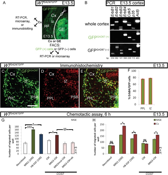
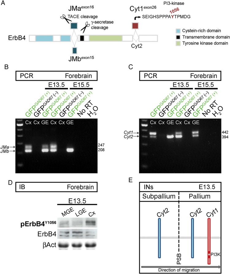
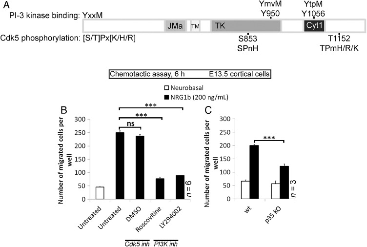
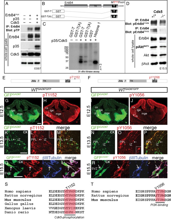
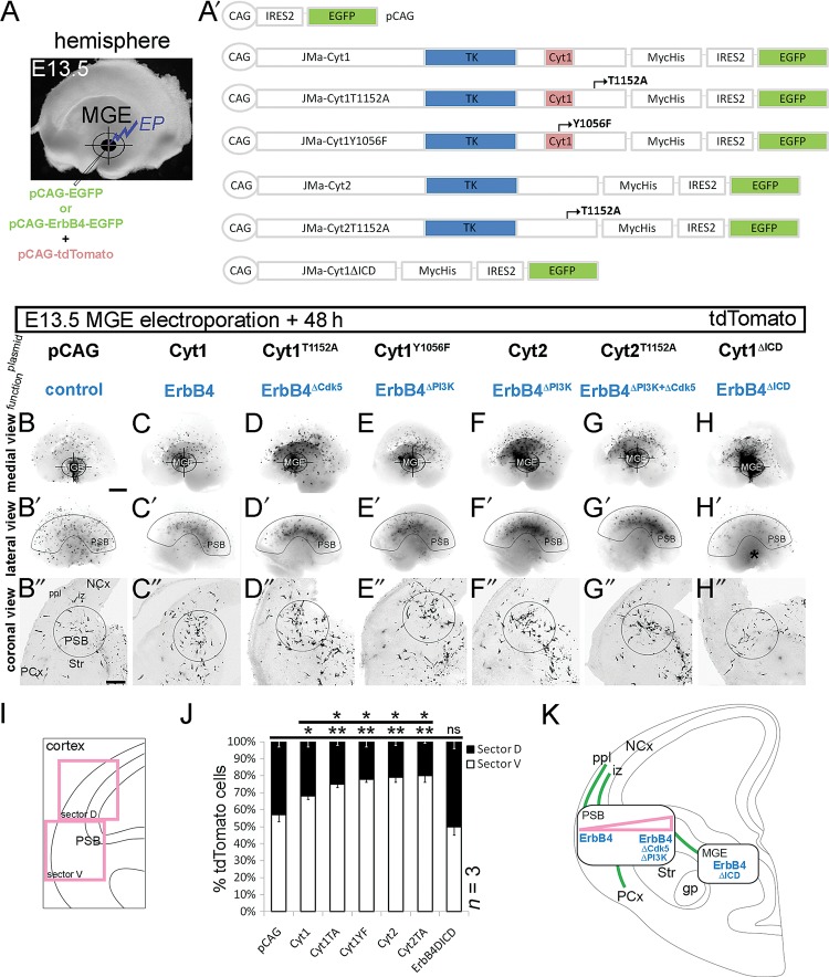
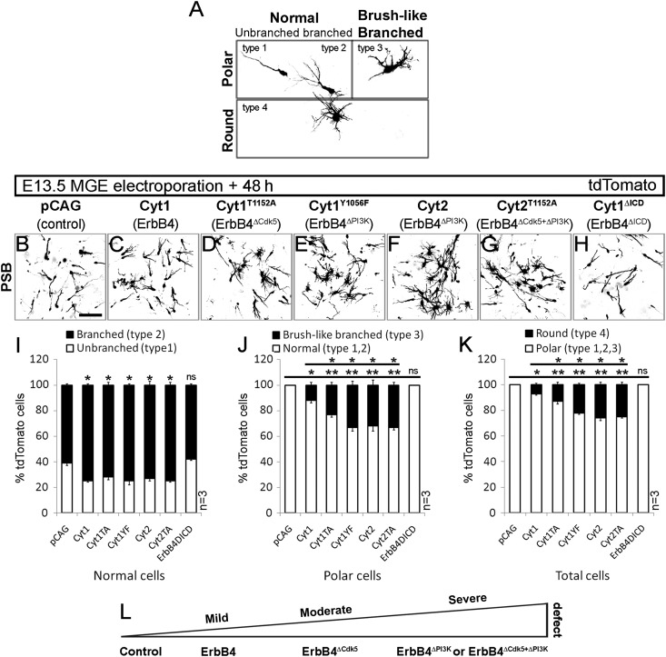
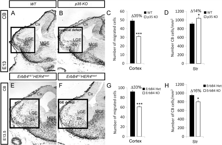
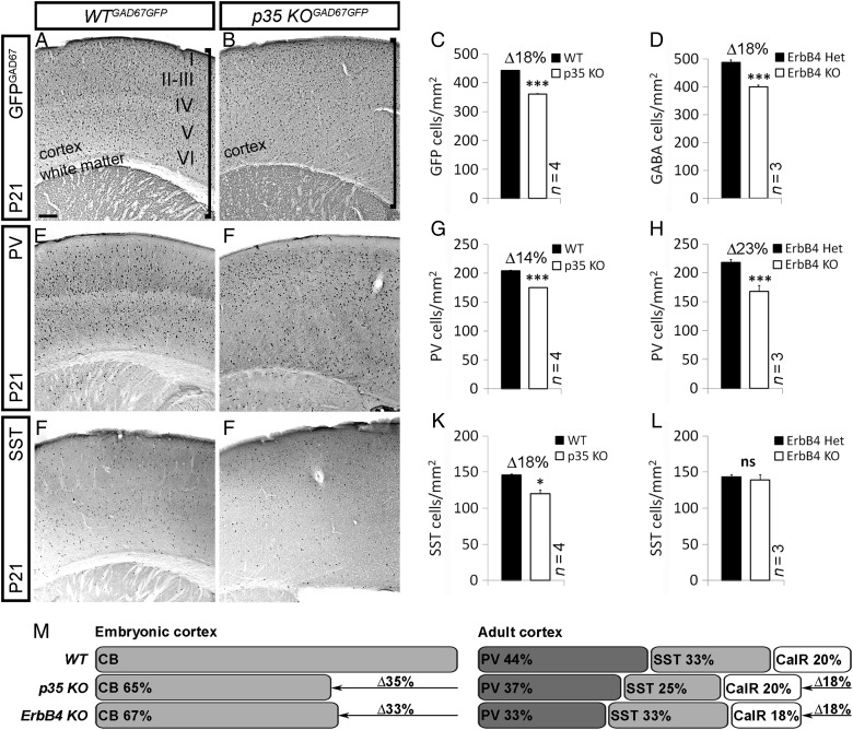
Interneuron dysfunction in humans is often associated with neurological and psychiatric disorders, such as epilepsy, schizophrenia, and autism. Some of these disorders are believed to emerge during brain formation, at the time of interneuron specification, migration, and synapse formation. Here, using a mouse model and a host of histological and molecular biological techniques, we report that the signaling molecule cyclin-dependent kinase 5 (Cdk5), and its activator p35, control the tangential migration of interneurons toward and within the cerebral cortex by modulating the critical neurodevelopmental signaling pathway, ErbB4/phosphatidylinositol 3-kinase, that has been repeatedly linked to schizophrenia. This finding identifies Cdk5 as a crucial signaling factor in cortical interneuron development in mammals.

Keywords: cerebral cortex; interneurons; migration; mouse; phosphorylation.


Figures

Figure 1.
Figure 1.
ErbB4 and its ligands facilitate migration of cortical interneurons in vitro. (A) Forebrain section of a GAD67GFP mouse embryo, showing areas/cells used in indicated experiments. GFPGAD67(+) cells represent GABAergic interneurons. (B) RT-PCR. ErbBs, p35, and Cdk5 expression in the developing Cx. Actβ is used as an internal control. (CF) Expression of ErbB4 in GFPGAD67(+) cells in the cortical migratory streams. (G, H) Chemotactic response of MGE- and Cx-derived cells to control (white) or to EGF-like domain of ErbB4 ligands: NRG1β (green), HB-EGF (blue), or NRG3-CM (burgundy). Negative controls for NRG3 (pink): NRG3mut-CM or NRG3-CM treated with a blocking NRG3 ab; (G) MGE cells; (H) MGE versus Cx cells. *P ≤ 0.05, **P ≤ 0.01, ***P ≤ 0.005, t-test. CM, conditioned medium. Bar, 50 μm.
Figure 2.
Figure 2.
Cortical interneurons upregulate Cyt1 expression in the pallium. (A) Schematic of ErbB4, illustrating domains and isoforms of the receptor (modified from Sundvall et al. 2008). JMa isoform, but not JMb, is susceptible to proteolytic cleavage. Cyt1 isoform contains a unique tyrosine residue (Y1056), absent in Cyt2, which serves as a binding site for PI3-kinase. (B, C) RT-PCR. ErbB4 isoform expression in FACS-purified forebrain cells from GAD67GFP mice at the indicated time points. GFPGAD67(+) cells represent GABAergic interneurons. (D) Immunoblots of protein lysates from the MGE, LGE, and Cx of E13.5 mice, showing phosphorylation (p) of ErbB4 on Y1056. ErbB4 and βAct serve as loading controls. (E) Schematic, illustrating upregulation of Cyt1 as early-born interneurons (INs) depart from the subpallium, cross the PSB and enter the pallium. Cyt, cytoplasmic; JM, juxtamembrane; RT, reverse transcriptase; TACE, tumor necrosis factor-α converting enzyme.
Figure 3.
Figure 3.
PI3-kinase and p35/Cdk5 pathways regulate ErbB4-mediated chemotaxis of cortical cells in vitro. (A) Scansite software, predicting PI3-kinase-binding and Cdk5-targeted phosphorylation sites of ErbB4. (B) Chemotactic response of Cx-derived cells, untreated or treated simultaneously with DMSO, Roscovitine (10 μM) or LY294002 (10 μM) for 30 min prior to assay, to NRG1β. (C) Chemotactic response of Cx-derived cells, wt or p35-KO, to NRG1β. *P ≤ 0.05, ***P ≤ 0.005, t-test.
Figure 4.
Figure 4.
p35/Cdk5 phosphorylation of ErbB4 in vitro and in vivo. (A) Immunoprecipitation (IP) of ErbB4myc from COS7 cell protein lysates; COS7 cells were co-transfected with ErbB4myc, p35, and Cdk5. Immunoblots of ErbB4 IP protein extracts, revealing p35/Cdk5 phosphorylation of ErbB4 on threonine adjacent to proline (pTP; first and second lanes vs. third lane). Immunoblots of whole-cell protein lysates indicate expression of ErbB4, p35, and Cdk5. βAct serves as a loading control. (B) Schematic, illustrating GST-fusion ErbB4 fragments used in a kinase assay. (C) Kinase assay, showing phosphorylation of ErbB4 on T1152 by p35/Cdk5. GST and histone 1 serve as negative and positive controls, respectively. (D) IP of ErbB4. Immunoblots of ErbB4 IP protein extracts and whole-cell protein lysates from the Cx of E15.5 mice, revealing decreased phosphorylation of ErbB4T1152, ErbB4Y1056, and AktS473 in the Cdk5 KOs compared with littermate controls. ErbB4, Akt, and βAct serve as loading controls. (E, F) Schematic diagrams of ErbB4, showing Cdk5-targeted (T1152; E) and PI3-kinase-binding (Y1056; F) phosphorylation sites recognized by pErbB4T1152 and pErbB4Y1056 antibodies, respectively. (GR) Forebrain sections of a GAD67GFP mouse embryo, at the indicated time points, immunostained for pErbB4T1152 and pErbB4Y1056 (red). GFPGAD67(+) cells (green) represent GABAergic interneurons. (GJ) Dotted lines indicate the border between the pallium (p) and the subpallium (s). (KP) show pErbB4T1152 and pErbB4Y1056 in a subset of cortical interneurons (arrows). Higher magnifications are shown in the insets. (Q) and (R) show that pErbB4T1152 and pErbB4Y1056 co-localize with β-III-tubulin (blue) in the proximal leading process (arrows) of cortical interneurons. (S,T) Cdk5-phosphorylation and PI3K-binding sites in ErbB4 are well conserved across species. Bars, 200 μm (FI), 50 μm (JO), 20 μm (P,Q). A, alanine; GST, glutathione S-transferase; SVZ, subventricular zone; Y, tyrosine.
Figure 5.
Figure 5.
Different ErbB4 signaling pathways regulate tangential distribution of MGE cells. (A) Focal electroporation (EP) of control (pCAG) vector and ErbB4 constructs (expressed from pCAG; A′), mixed with a CAG-driven tdTomato vector, into the E13.5 MGE of the whole-mouse telencephalic hemisphere. (BH″) Fluorescent images of tdTomato, converted to grayscale mode, 48-h post electroporation. (BH) Medial view of the telencephalic hemisphere. Target symbol specifies the site of electroporation (MGE). (B′–H′) Lateral view of the telencephalic hemisphere, depicting the extent of tangential cell spread. The arcuate-shaped outline indicates the position of the PSB. (H′) Asterisk, MGE. (B″–H″) Coronal view of the sectioned telencephalic hemisphere, showing the cortical migratory streams. A circle outlines the PSB. (I, J) Quantification of the distribution of MGE cells in the Cx. (I) Schematic, showing the Cx divided into 2 equal sectors, ventral (marked “V,” which includes the PSB) and dorsal (marked “D”). (J) A graph, showing the result of quantification. *P ≤ 0.05, **P ≤ 0.01, t-test comparing with pCAG. (K) Schematic representations of EP experiments: green lines depict the migratory routes of interneurons, rectangles indicate the sites of abnormal accumulation of cells overexpressing ErbB4 plasmids (blue), and triangle (pink) signifies the severity of the defect. gp, globus pallidus; NCx, neocortex; PCx, paleocortex; Str, striatum. Bar, 250 μm.
Figure 6.
Figure 6.
Leading process morphology defects of cortical interneurons after loss of Cdk5 and PI3-kinase ErbB4 signaling. (AH) tdTomato fluorescence, converted to grayscale, revealing morphology of migrating interneurons, 48-h postelectroporation of E13.5 MGE with control (pCAG; B) and ErbB4 (CH) constructs (see Fig. 5). (A) Four types of MGE cell-leading process morphologies, observed at the PSB. (I) Percentage of unbranched (type 1) and branched (type 2) cells relative to total number of normal (type 1, 2) cells. (J) Percentage of brush-like branched (type 3) and normal (type 1, 2) cells relative to total number of polar (type 1, 2, and 3) cells. (K) Percentage of round (type 4) and polar (type 1, 2, and 3) cells relative to total cell number (type 1, 2, 3, and 4). (L) Schematic, indicating the severity of morphology defect in MGE cells in respect to altered ErbB4 signaling pathway. *P ≤ 0.05 (mild), **P ≤ 0.01 (moderate), ***P ≤ 0.005 (severe), t-test comparing against either control or Cyt1. Bar, 50 μm.
Figure 7.
Figure 7.
Loss of p35 and ErbB4 alter the number of interneurons that reach the Cx via different mechanisms. (A, B, E, F) Embryonic forebrain sections of indicated genotypes and ages, immunostained for CB. The LGE/Cx junction is shown within a box outline. (C, D, G, F) Quantification of the number of CB cells in the Cx and striatum (Str) of control (black) and indicated KO (white) animals. *P ≤ 0.05, ***P ≤ 0.005, t-test. Bar, 200 μm.
Figure 8.
Figure 8.
Loss of p35 and ErbB4 results in reduction of a subset of interneurons in the adult Cx. (A, B, E, F, I, J) Sections of adult (P21) somatosensory Cx of control and p35 KO mouse, immunostained for GFP, PV, and SST, indicating the quantity and distribution of interneurons. (A, B) Vertical line specifies the width of the Cx and white matter. Roman numerals designate cortical layers in control animals. Loss of p35 alters cortical laminar organization (layers are inverted compared with controls), and white matter is histologically undetectable due to misguided fibers that run through the center of the Cx (Rakić et al. 2006). (C, D, G, H, K, L) Quantification of the number of GFPGAD67, PV, and SST interneurons in the Cx of control (black) and indicated KO (white) animals. *P ≤ 0.05, ***P ≤ 0.005, t-test. (M) Schematic, showing loss of embryonic and postnatal interneurons in p35 and ErbB4 KOs compared with controls. Bar, 50 μm.

References

    1. Ang ES, Jr, Haydar TF, Gluncic V, Rakic P. Four-dimensional migratory coordinates of GABAergic interneurons in the developing mouse cortex. J Neurosci. 2003;23:5805–5815. - PMC - PubMed
    1. Anton ES, Ghashghaei HT, Weber JL, McCann C, Fischer TM, Cheung ID, Gassmann M, Messing A, Klein R, Schwab MH, et al. Receptor tyrosine kinase ErbB4 modulates neuroblast migration and placement in the adult forebrain. Nat Neurosci. 2004;7:1319–1328. doi: 10.1038/nn1345. - DOI - PubMed
    1. Assimacopoulos S, Grove EA, Ragsdale CW. Identification of a Pax6-dependent epidermal growth factor family signaling source at the lateral edge of the embryonic cerebral cortex. J Neurosci. 2003;23:6399–6403. - PMC - PubMed
    1. Baudoin JP, Alvarez C, Gaspar P, Métin C. Nocodazole-induced changes in microtubule dynamics impair the morphology and directionality of migrating medial ganglionic eminence cells. Dev Neurosci. 2008;30:132–143. doi: 10.1159/000109858. - DOI - PubMed
    1. Bellion A, Baudoin JP, Alvarez C, Bornens M, Métin C. Nucleokinesis in tangentially migrating neurons comprises two alternating phases: forward migration of the Golgi/centrosome associated with centrosome splitting and myosin contraction at the rear. J Neurosci. 2005;25:5691–5699. doi: 10.1523/JNEUROSCI.1030-05.2005. - DOI - PMC - PubMed

Publication types

MeSH terms