. 2014 Mar;99(3):465-73.
doi: 10.3324/haematol.2013.093765. Epub 2013 Oct 18.

Next-generation sequencing-based multigene mutational screening for acute myeloid leukemia using MiSeq: applicability for diagnostics and disease monitoring

Affiliations

Next-generation sequencing-based multigene mutational screening for acute myeloid leukemia using MiSeq: applicability for diagnostics and disease monitoring

Rajyalakshmi Luthra et al. Haematologica. 2014 Mar.

Abstract

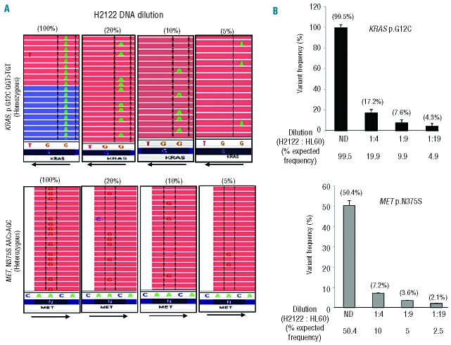
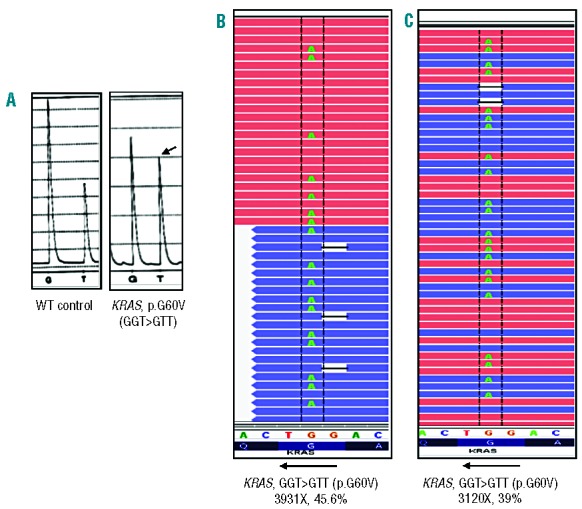
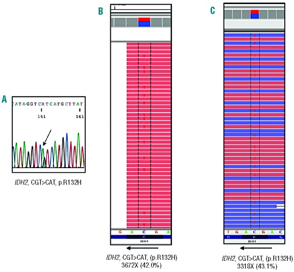
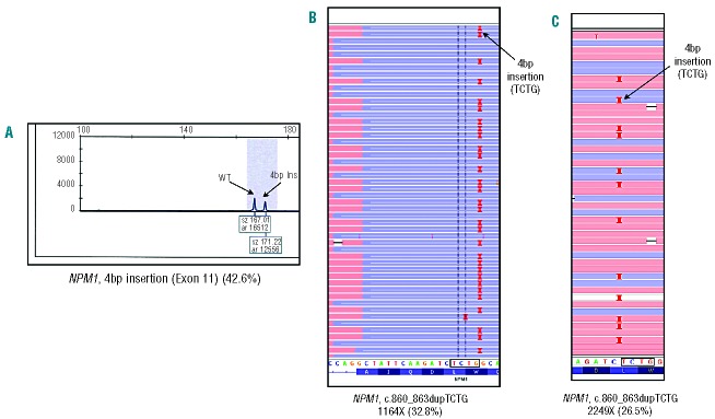
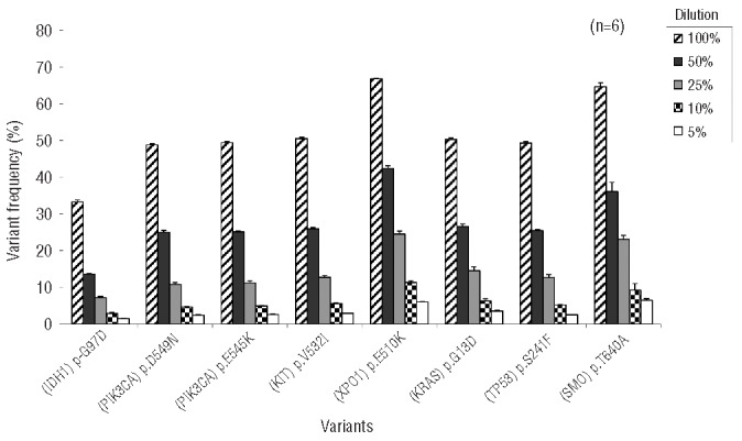
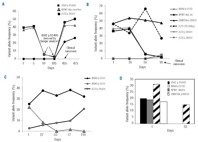
Routine molecular testing in acute myeloid leukemia involves screening several genes of therapeutic and prognostic significance for mutations. A comprehensive analysis using single-gene assays requires large amounts of DNA, is cumbersome and timely consolidation of results for clinical reporting is challenging. High throughput, next-generation sequencing platforms widely used in research have not been tested vigorously for clinical application. Here we describe the clinical application of MiSeq, a next-generation sequencing platform to screen mutational hotspots in 54 cancer-related genes including genes relevant in acute myeloid leukemia (NRAS, KRAS, FLT3, NPM1, DNMT3A, IDH1/2, JAK2, KIT and EZH2). We sequenced 63 samples from patients with acute myeloid leukemia/myelodysplastic syndrome using MiSeq and compared the results with those obtained using another next-generation sequencing platform, Ion-Torrent Personal Genome Machine and other conventional testing platforms. MiSeq detected a total of 100 single nucleotide variants and 23 NPM1 insertions that were confirmed by Ion Torrent or conventional platforms, indicating complete concordance. FLT3-internal tandem duplications (n=10) were not detected; however, re-analysis of the MiSeq output by Pindel, an indel detection algorithm, did detect them. Dilution studies of cancer cell-line DNA showed that the quantitative accuracy of mutation detection was up to an allelic frequency of 1.5% with a high level of inter- and intra-run assay reproducibility, suggesting potential utility for monitoring response to therapy, clonal heterogeneity and evolution. Examples demonstrating the advantages of MiSeq over conventional platforms for disease monitoring are provided. Easy work-flow, high throughput multiplexing capability, 4-day turnaround time and simultaneous assessment of routinely tested and emerging markers make MiSeq highly applicable for clinical molecular testing in acute myeloid leukemia.


Figures

Figure 1.
Figure 1.
Sensitivity of detection using MiSeq. (A) A representative image of the aligned sequencing reads as visualized in Integrative Genome Viewer shows a progressive decrease in the single nucleotide variant detected in the background of wild-type sequence in H2122 cell line DNA sequentially diluted into HL60 cell line DNA. With dilution, a clear and proportional decrease in the homozygous KRAS (GGT>TGT, p.G12C) and a heterozygous MET (AAC>AGC, p.N375S) mutation are evident. KRAS gene has the reverse orientation on chromosome 12. However, by default, the Integrative Genome Viewer exhibits aligned reads in ‘forward’ orientation. Hence the substituted nucleotide appears as ‘A’ instead of ‘T’ in the reads (CCA>ACA or GGT>TGT). The directions of gene orientation are indicated by the arrows. (B) The expected variant frequencies and the average variant frequency of the KRAS (p.G12C) and MET (p.N375S) mutations detected in two independent sensitivity analyses.
Figure 2.
Figure 2.
Concordance of MiSeq, pyrosequencing and Ion Torrent Personal Genome Machine (IT-PGM) findings. A representative sample in which a KRAS mutation (p.G60V) identified by pyrosequencing was also detected and called by both MiSeq and the IT-PGM. (A) The pyrosequencing results showing the base change (GGT>GTT, indicated by the arrow) in KRAS in comparison with wild-type control. (B) The same mutation change is evident in the aligned reads from the MiSeq sequencing output at a very high coverage of 3,931X and showing a variant frequency of 45.6%. (C) Sequencing on the IT-PGM also confirms the same mutation at a sequencing depth and variant frequency comparable with the MiSeq. The KRAS gene has the reverse orientation on chromosome 12. As the default setting on Integrative Genome Viewer exhibits aligned reads in ‘forward’ orientation, the substituted nucleotide appears as ‘A’ instead of ‘T’ in the reads (CCA>CAA or GGT>GTT). The orientation of the gene is depicted by the arrows in panels (B) and (C).
Figure 3.
Figure 3.
Concordance of MiSeq, Sanger sequencing and Ion Torrent Personal Genome Machine (IT-PGM). A mutation in IDH2 (p.R132H) resulting from a CGT>CAT substitution originally detected by Sanger sequencing (A) is also detected clearly in the MiSeq sequencing output and (B) sequencing using the IT-PGM with comparable coverage and mutational frequency with both platforms. IDH2 has reverse orientation on chromosome 15. The default setting on Integrative Genome Viewer shows aligned reads in forward orientation only. Hence, the substituted nucleotide appears as ‘T’ instead of ‘A’ in the reads (CGT>CAT or GCA>GTA). The orientation of the gene is depicted by the arrows in panels (B) and (C).
Figure 4.
Figure 4.
Ability of sequencing using MiSeq to detect insertions in NPM1. (A) A characteristic 4 bp insertion (G>GTCTG) in exon 12 of the NPM1 gene is evident in this sample as detected by capillary electrophoresis and is present at a ratio of 0.426 or 42.6%. (B) Sequencing of the same sample using MiSeq also detected and called this mutation at a sequencing depth of 1,164X and a variant frequency of 32.8%. (C) The Ion Torrent Personal Genome Machine (IT-PGM) also detected and called the same 4 bp insertion at a sequencing depth of 2,249X and a variant frequency of 26.5% in the background of wild-type sequence. The presence of the insertions in the aligned reads is indicated as characteristic red bars in Integrative Genome Viewer as seen in panels (B) and (C) (indicated by the arrows).
Figure 5.
Figure 5.
Assay sensitivity of the V2 upgrade. Undiluted (100%) and samples of DLD1 cell line DNA sequentially diluted into normal female control DNA (50%, 25%, 10% and 5%) were sequenced in six different sequencing runs and the ability to detect the eight different heterozygous (monoallelic) mutations present in each dilution was tested. These mutations were successfully detected in every dilution tested in all of the sequencing runs with minimal variation in the variant frequency detected.
Figure 6.
Figure 6.
Application for disease monitoring. (A) IDH2 and NPM1 mutations detected in a patient at the onset of the disease were missed subsequently when present at low levels (days 10 and 185) when monitored by Sanger sequencing but were effectively detected and called by MiSeq. The time point of clinical remission is indicated by the arrow (B) Mutations in DNMT3A and NRAS, and insertions in NPM1 and FLT3 were detected at diagnosis and followed subsequently. Two low-lying FLT3 D835 mutations appeared at day 95 indicating the need to follow multiple mutations. The FLT3 ITD percentages as detected by capillary electrophoresis are plotted. The time point of clinical remission is indicated by the arrow (C) Two NRAS mutations were detected, one of which disappeared with treatment indicating distinct clones. A FLT3 Y842H activating mutation, not routinely tested for was also detected by multigene screening on MiSeq (D) Mutations were detected in four genes at diagnosis warranting testing of each of them after treatment. The mutiplexing capacity of MiSeq would help in consolidating them on to a single platform. Data presented in panels (C) and (D) were from patients with refractory AML.

Similar articles

Cited by

References

    1. Owen C, Barnett M, Fitzgibbon J. Familial myelodysplasia and acute myeloid leukaemia a review. Br J Haematol. 2008;140(2):123–32 - PubMed
    1. Sanders MA, Valk PJ. The evolving molecular genetic landscape in acute myeloid leukaemia. Curr Opin Hematol. 2013;20(2): 79–85 - PubMed
    1. Ley TJ, Ding L, Walter MJ, McLellan MD, Lamprecht T, Larson DE, et al. DNMT3A mutations in acute myeloid leukemia. N Engl J Med. 2010;363(25):2424–33 - PMC - PubMed
    1. Ley TJ, Mardis ER, Ding L, Fulton B, McLellan MD, Chen K, et al. DNA sequencing of a cytogenetically normal acute myeloid leukaemia genome. Nature. 2008; 456(7218):66–72 - PMC - PubMed
    1. Ding L, Ley TJ, Larson DE, Miller CA, Koboldt DC, Welch JS, et al. Clonal evolution in relapsed acute myeloid leukaemia revealed by whole-genome sequencing. Nature. 2012;481(7382):506–10 - PMC - PubMed

MeSH terms