. 2013 Oct 15;8(10):e78105.
doi: 10.1371/journal.pone.0078105. eCollection 2013.

The mitochondrial RNA landscape of Saccharomyces cerevisiae

Affiliations

The mitochondrial RNA landscape of Saccharomyces cerevisiae

Edward M Turk et al. PLoS One. .

Abstract

Mitochondria are essential organelles that harbor a reduced genome, and expression of that genome requires regulated metabolism of its transcriptome by nuclear-encoded proteins. Despite extensive investigation, a comprehensive map of the yeast mitochondrial transcriptome has not been developed and all of the RNA-metabolizing proteins have not been identified, both of which are prerequisites to elucidating the basic RNA biology of mitochondria. Here, we present a mitochondrial transcriptome map of the yeast S288C reference strain. Using RNAseq and bioinformatics, we show the expression level of all transcripts, revise all promoter, origin of replication, and tRNA annotations, and demonstrate for the first time the existence of alternative splicing, mirror RNAs, and a novel RNA processing site in yeast mitochondria. The transcriptome map has revealed new aspects of mitochondrial RNA biology and we expect it will serve as a valuable resource. As a complement to the map, we present our compilation of all known yeast nuclear-encoded ribonucleases (RNases), and a screen of this dataset for those that are imported into mitochondria. We sought to identify RNases that are refractory to recovery in traditional mitochondrial screens due to an essential function or eclipsed accumulation in another cellular compartment. Using this in silico approach, the essential RNase of the nuclear and cytoplasmic exosome, Dis3p, emerges as a strong candidate. Bioinformatics and in vivo analyses show that Dis3p has a conserved and functional mitochondrial-targeting signal (MTS). A clean and marker-less chromosomal deletion of the Dis3p MTS results in a defect in the decay of intron and mirror RNAs, thus revealing a role for Dis3p in mitochondrial RNA decay.


Conflict of interest statement

Competing Interests: The authors have declared that no competing interests exist.

Figures

Figure 1
Figure 1. The transcriptome of yeast S288C mitochondria.
(A) Physical, genetic, and transcriptome map of the yeast S288C mitochondrial genome. The positive strand is on the outside, and the negative is inside. The active ori (green arrows), inactive ori (grey arrows), primary transcripts (blue arrows), dodecamers (red lollipops), and deviant dodecamers (yellow lollipops) are modeled. The location of each gene is depicted using the systematic name, protein-coding genes are uppercase/bold, and the transcription start sites are numbered. To mark genes within introns, the intron is lowercase/first, “+”, and then the systematic name of the gene (B) The steady-state RNA abundance of all mitochondrial-encoded genes and introns measured in RPKM.
Figure 2
Figure 2. The ori of yeast S288C mitochondria.
(A) Model based on the presumably active ori2-3-5, depicting the non-template strand. The structure consists of a 20-bp promoter (invariable length), followed by a 17-bp G-rich sequence (invariable length and sequence), then an A/T-rich spacer of ~200 bp, followed by two complementary 7-bp sequences (invariable length and sequence). The 7-bp sequences are separated by a 29-bp A/T-rich sequence (invariable length). (B) Alignment of the 20-bp promoters of the potentially active (2-3-5) and inactive (1-4-6-7-8) ori. Single nucleotide differences for ori3 and 4 are highlighted in red, and GC-elements that disrupt the promoter are in green.
Figure 3
Figure 3. Alternative splicing of aI5β.
(A) Model of the 13 introns of yeast S288C mitochondria, and 10 HEG-related ORFs contained within them. The first five introns of COX1, and three COB introns, contain ORFs that are in-frame with their upstream exon. The ORF within the omega intron of 21S rRNA encodes an ATG at the 5’ end and a dodecamer at the 3’ end. The sixth intron of COX1, aI5β, contains an intron that is not in-frame with the upstream exon and does not encode an ATG at the 5’ end. (B) Electropherogram of the sequenced RT-PCR product derived from alternatively spliced aI5β. For comparison, the sequence derived from primary splicing or alternative splicing is depicted above the electropherogram. In both cases the 5’ exon is the sixth COX1 exon. The 3’ exon is the seventh COX1 exon in the case of primary splicing and the ORF within the aI5β intron in the case of alternative splicing.
Figure 4
Figure 4. The polypeptide-coding genes of yeast S288C mitochondria.
Each gene is labeled using the standard name (www.yeastgenome.org) and convention (all capital, italic, no spaces), or the systematic name (all capital, regular) if no standard name is available. Each open reading frame begins with the AUG codon (green) and ends with the UAA stop codon (red). The size of the ORF in between the start and stop codons is depicted as number of bases, as is the distance in between the stop codon and the dodecamer (12 mer) sequence, which is blue with deviations bold/black. The likely cleavage sites are depicted with a down arrow. The BI3 and SCEI stop codons (bold/red) are within the dodecamer. The heptakaidecamer (17 mer) downstream of the ATP8 and BI3 dodecamer is underlined. The distance in between the OLI1 / ATP9 dodecamer and the downstream tRNA (Ser) that cleaves the primary transcript via normal tRNA processing is depicted as number of bases.
Figure 5
Figure 5. Heptakaidecamer cleavage site.
(A) Alignment of four heptakaidecamer sequences. The down arrow that separates the blue and the orange sequences indicates the cleavage site. (B) The non-template strand of the 483-bp, unprocessed, RNA subunit of RNase P, derived from the mitochondrial-encoded RPM1 gene. Boxes represent the three fragments identified in purified RNase P. Fragment 1 (89 nt) is at the 5’ end of the primary transcript, and fragments 2 (71 nt) and #3 (52 nt) overlap and are positioned at the 3’ end. The 5’ fragment is separated from the 3’ fragments by a heptakaidecamer cleavage site highlighted in blue and orange. The base immediately upstream and downstream of cleavage is underlined.
Figure 6
Figure 6. Dis3p is a conserved mitochondrial protein.
(A) I-TASSER models of Dis3p and Arg8p. N-termini indicated by arrows. (B) Amphipathic alpha helix located at the N-terminus of Dis3p and Arg8p. Single letter amino acid code and position in the primary structure is indicated. (C) Growth of yeast on arginine-free medium when the MTS of DIS3 replaces the plasmid borne MTS of ARG8. (D) The Drosophila dDis3 N-terminus can function as an MTS for GFP, but mutation of the MTS (4A) cannot. (E) Western blot of cell fractionation. C (cytoplasm), M (mitochondria).
Figure 7
Figure 7. Top 50 alignments to yeast Dis3p shows a strongly conserved N-terminus.
Species name is followed by NCBI reference, the amino acid sequence of the N-terminus, and the percent likelihood of mitochondrial targeting. Down arrow marks the conserved mitochondrial cleavage site.
Figure 8
Figure 8. Engineering scheme to create dis3∆mts.
(A) Amplified the CORE molecule by PCR from plasmid pGSKU using primers 9H9 / 9H10, which include targeting sequences at their 5‘ ends. (B) Amplified the upstream targeting molecule by PCR from yeast chromosomal DNA, strain BY4741, using primers 9C7 / 9F3. Amplified the downstream targeting molecule by PCR from yeast chromosomal DNA, strain P CUP1 -DIS3 which is a derivative of BY4741, using primers 9I1 / 8G9. (C) Simultaneously transformed the three molecules into BY4741. (D) Transformation resulted in a strain in which the MTS is deleted, DIS3 is expressed by the CUP1 promoter, and identical self-cloning sites (SCS) flank the cassette. (E) Over-night growth in galactose media induced expression of SCE1 endonuclease, which cleaved the DNA at the restriction site (RS), and the cut was repaired by homologous recombination between the SCS, which removed the cassette. The entire gene was amplified by PCR using primers 9E10 / 9F1 and sequenced with the PCR primers and internal primers 8G8, 9C8, and 9D3.
Figure 9
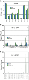
Figure 9. The mRNA and mirror-RNA profiles in DIS3 and dis3∆mts.
Wild type (DIS3) reads are first / blue and the mutant (dis3∆mts) are second / green. Error bars = s.e.m. Asterisk denotes a statistically significant difference with a p-value less than 0.01 as determined by the DESeq statistical method[29]. (A) Read count in RPKM for the eight mRNAs that encode proteins required for oxidative phosphorylation. (B) Total number of reads that map to each mirror ORF. (C) Total number of reads that map to each mirror tRNA.
Figure 10
Figure 10. Hyper-accumulation of Group I and Group II intron RNA in dis3∆mts.
(A) Fold change of the RPKM value between DIS3 and dis3∆mts for the exons and introns of 21S rRNA, COX1, and COB. Values >1 indicate an increase in reads in dis3∆mts. Only the first and last exon (orange) of each intron-containing gene is shown for purposes of clarity. Error bars = s.e.m. Asterisk denotes a statistically significant difference with a p-value less than 0.01 as determined by the DESeq statistical method[29]. (B) RT-qPCR of DIS3 and dis3∆mts RNA. Change in Ct value for the first exon of 21S rRNA (orange), the first and last exon of COB (orange), the five COB introns (blue), and ligated exons (LE / green) of the COB gene. Values greater than zero indicate greater abundance in dis3∆mts. Error bars = s.e.m. Asterisk denotes a statistically significant difference with a p-value less than 0.01 as determined by the Student’s t-test.

References

    1. Ephrussi B (1949) Action de l’acriflavine sur les levures. In: Unités Biologiques Douées de Continuité Génétiqué. du Paris: Centre Nat Rech Sci: 165-180.
    1. Hagemann R (2010) The foundation of extranuclear inheritance: plastid and mitochondrial genetics. Mol Genet Genomics 283: 199-209. doi:10.1007/s00438-010-0521-z. PubMed: 20140454. - DOI - PubMed
    1. Goffeau A, Barrell BG, Bussey H, Davis RW, Dujon B et al. (1996) Life with 6000 genes. Science 274: 546: 563-547. PubMed: 8849441. - PubMed
    1. Goffeau A, Aert R, Agostini-Carbone ML, Ahmed A, Aigle M (1997) The yeast genome directory. Nature 387: 5. doi:10.1038/387005b0. PubMed: 9169864. - DOI - PubMed
    1. Foury F, Roganti T, Lecrenier N, Purnelle B (1998) The complete sequence of the mitochondrial genome of Saccharomyces cerevisiae. FEBS Lett 440: 325-331. doi:10.1016/S0014-5793(98)01467-7. PubMed: 9872396. - DOI - PubMed

Publication types

MeSH terms