. 2013 Jul 3;2(9):891-900.
doi: 10.1242/bio.20133632. eCollection 2013.

Metastatic MTLn3 and non-metastatic MTC adenocarcinoma cells can be differentiated by Pseudomonas aeruginosa

Affiliations

Metastatic MTLn3 and non-metastatic MTC adenocarcinoma cells can be differentiated by Pseudomonas aeruginosa

Matthew J Novotny et al. Biol Open. .

Abstract

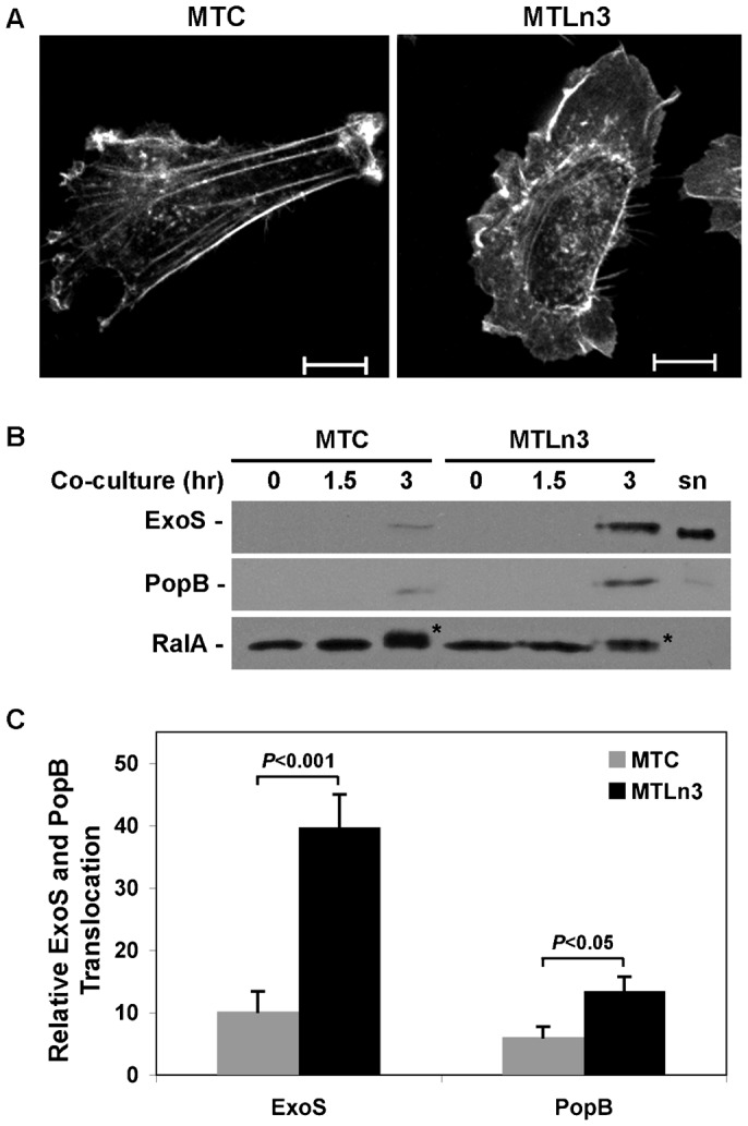
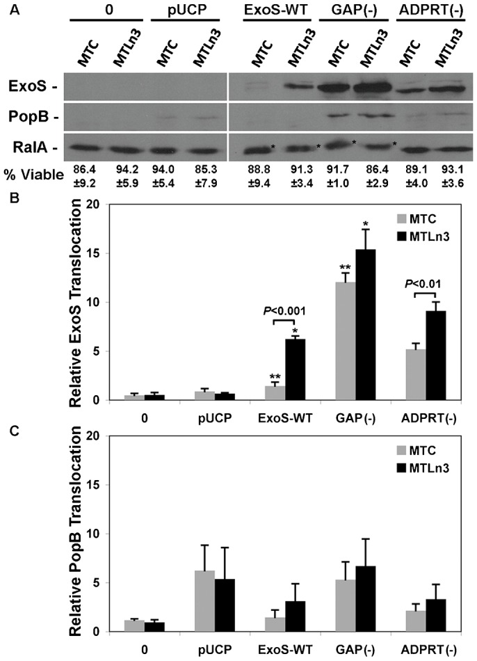
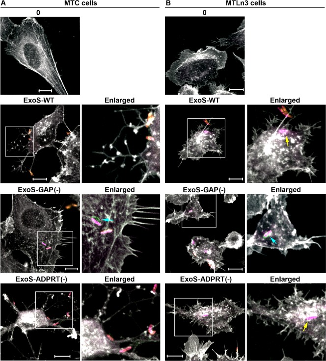
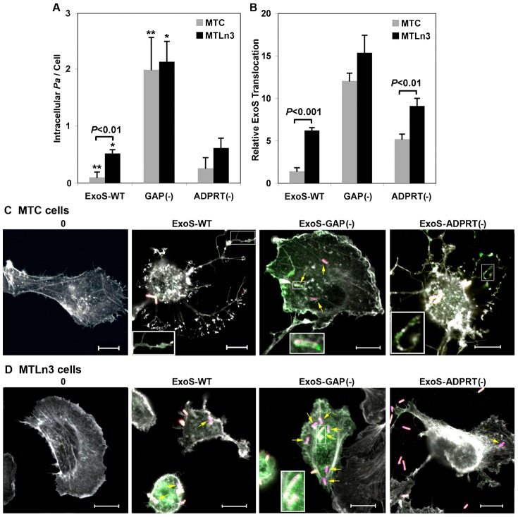
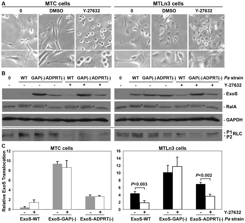
Cancer patients are known to be highly susceptible to Pseudomonas aeruginosa (Pa) infection, but it remains unknown whether alterations at the tumor cell level can contribute to infection. This study explored how cellular changes associated with tumor metastasis influence Pa infection using highly metastatic MTLn3 cells and non-metastatic MTC cells as cell culture models. MTLn3 cells were found to be more sensitive to Pa infection than MTC cells based on increased translocation of the type III secretion effector, ExoS, into MTLn3 cells. Subsequent studies found that higher levels of ExoS translocation into MTLn3 cells related to Pa entry and secretion of ExoS within MTLn3 cells, rather than conventional ExoS translocation by external Pa. ExoS includes both Rho GTPase activating protein (GAP) and ADP-ribosyltransferase (ADPRT) enzyme activities, and differences in MTLn3 and MTC cell responsiveness to ExoS were found to relate to the targeting of ExoS-GAP activity to Rho GTPases. MTLn3 cell migration is mediated by RhoA activation at the leading edge, and inhibition of RhoA activity decreased ExoS translocation into MTLn3 cells to levels similar to those of MTC cells. The ability of Pa to be internalized and transfer ExoS more efficiently in association with Rho activation during tumor metastasis confirms that alterations in cell migration that occur in conjunction with tumor metastasis contribute to Pa infection in cancer patients. This study also raises the possibility that Pa might serve as a biological tool for dissecting or detecting cellular alterations associated with tumor metastasis.

Keywords: Cell migration; MTC and MTLn3 cells; Pseudomonas aeruginosa; Rho GTPase; Tumor metastasis.


Conflict of interest statement

Competing interests: The authors have no competing interests to declare.

Figures

Fig. 1.
Fig. 1.. Highly metastatic MTLn3 cells exhibit a different migratory phenotype and allow greater Pa-T3S translocation than non-metastatic MTC cells.
(A) MTC and MTLn3 cell morphology was compared under steady state conditions (5% FBS) in the absence of Pa. Cells were stained with Phalloidin-TRITC to detect actin (white). Scale bar: 10 µm. (B) MTC and MTLn3 cells were co-cultured with strain PA103ΔUT expressing plasmid encoded HA tagged wild type ExoS (Pa ExoS-WT) for the indicated times. Cells were harvested, fractionated and T3S translocation of ExoS and PopB into membrane fractions was assayed by immunoblot analysis based on equal protein loading. RalA modification served as a functional read-out of translocated ExoS-ADPRT activity, and asterisks mark ADP-ribosylated RalA. Total RalA served as a membrane fraction loading control. (sn) T3S induced Pa ExoS-WT culture supernatant was used as a molecular marker for ExoS and PopB. (C) T3S translocated ExoS and PopB were quantified by densitometry, and significant differences in ExoS and PopB translocation between MTC and MTLn3 cells are indicated. Results represent the mean ± s.e.m. of five independent experiments.
Fig. 2.
Fig. 2.. ExoS-GAP activity determines differences in T3S translocation in MTC and MTLn3 cells.
(A) MTC and MTLn3 cells were co-cultured for 3 hr with Pa ExoS-WT, Pa expressing ExoS with a R146A GAP(−) mutant, or Pa expressing ExoS with an E379A and E381A ADPRT(−) mutant. Controls include uninfected cells (0) or cells co-cultured with a Pa pUCP plasmid control. Cells were harvested, fractionated and T3S translocation of ExoS and PopB into membrane fractions was assayed as in Fig. 1. (B) T3S translocated ExoS following co-culture with the indicated Pa strain was quantified by densitometry, and the mean ± s.e.m. of three independent experiments are represented. Significant differences between MTC and MTLn3 translocation of ExoS-WT and ExoS-ADPRT(−) are indicated. No significant difference in ExoS-GAP(−) translocation between MTC and MTLn3 cells was observed. Asterisks mark significant differences in ExoS-WT and ExoS-GAP(−) translocation into MTC cells (**P<0.001), or in ExoS-WT and ExoS-GAP(−) translocation into MTLn3 cells (*P<0.01). A significant (P<0.01) increase in translocation of ExoS-ADPRT(−) as compared to ExoS-WT was also detected in MTC cells, which is not indicated in Fig. 2B for simplicity. (C) T3S translocated PopB following co-culture with the indicated Pa strain was quantified by densitometry, and the mean ± s.e.m. of three independent experiments are represented. Differences observed in PopB translocation did not gain statistical significance.
Fig. 3.
Fig. 3.. ExoS-GAP activity differentially alters MTC and MTLn3 cell morphology.
(A) MTC or (B) MTLn3 cells were co-cultured for 1 hr 45 min with Pa expressing active GA, ExoS-WT or ExoS-ADPRT(−); inactive GAP, Pa ExoS-GAP(−); or with no Pa (0). Following co-culture cells were stained for extracellular Pa (yellow + pink), then fixed and permeabilized and stained for intracellular Pa (pink). ExoS effector was detected using an anti-HA antibody (green), and F-actin was stained using phalloidin (white). Scale bars: 10 µm. Regions of images enclosed in boxes were enlarged to allow better visualization of alterations in actin leading edge architecture following co-culture with Pa expressing the indicated ExoS effector. In enlarged images, intracellular Pa are indicated with yellow arrows, and intracellular Pa secreting ExoS effector are indicated with blue arrows. Results are representative of four independent experiments.
Fig. 4.
Fig. 4.. Metastatic properties of MTLn3 cells enhance Pa internalization.
(A) MTC and MTLn3 cells were co-cultured for 1 hr 45 min to 3 hr in four independent experiments with Pa ExoS-WT, Pa ExoS-GAP(−) or Pa ExoS-ADPRT(−). Cells were stained for extracellular or intracellular Pa as in Fig. 3, and Pa internalization was visually enumerated based on Pa association with an average of 140 MTC or MTLn3 cells per strain. Results are expressed as the mean ± s.e.m. of Pa internalized per MTC or MTLn3 cell. Significant difference (P<0.01) in Pa ExoS-WT internalization in MTC and MTLn3 is indicated. Asterisks mark significant differences in Pa ExoS-WT and Pa ExoS-GAP(−) internalization in MTC cells (**P<0.001) or Pa ExoS-WT and Pa ExoS-GAP(−) internalization in MTLn3 cells (*P<0.01). (B) Compares ExoS data shown in Fig. 2B with Pa internalization data (A). Significant differences are indicated. (C) MTC and (D) MTLn3 cells were uninfected (0) or co-cultured for 3 hr with Pa ExoS-WT, Pa ExoS-GAP(−) or Pa ExoS-ADPRT(−). Cells were stained as in Fig. 3 for extracellular Pa (yellow + pink), intracellular Pa (pink, indicated with yellow arrows), ExoS effector (green), and actin (white). Insets show enlargement of regions where ExoS effector co-localizes with actin (MTC cells: ExoS-WT or ExoS-ADPRT(−) images), or where ExoS effector co-localizes with intracellular Pa (ExoS-GAP(−): MTC and MTLn3 cell images). Scale bars: 10 µm. Results are representative of four independent experiments, but images from a single experiment are compared in panels C and D.
Fig. 5.
Fig. 5.. Inhibition of ROCK activity in MTLn3 cells decreased ExoS translocation.
(A) MTC and MTLn3 cell morphology was examined by phase contrast microscopy following no treatment (0), serum starvation for 3 hr and treatment for 30 min with DMSO (control), or serum starvation and treatment for 30 min with 25 µM Y-27632. MTC cells severely contracted in response to ROCK inhibitor, while MTLn3 cells adapted an elongated morphology with a severely contracted trailing edge. Images were taken at 20× magnification. (B) Following treatment with ROCK inhibitor, MTC and MTLn3 cells were either uninfected (0) or co-cultured for 3.5 hr with Pa ExoS-WT, Pa ExoS-GAP(−) or Pa ExoS-ADPRT(−) under steady state conditions. Cells were washed, detached with trypsin, resuspended in Laemmli buffer, and ExoS translocation was examined by SDS-11%PAGE analysis based on equal protein loading. RalA modification served as a functional read-out of translocated ExoS-ADPRT activity, and is indicated by an asterisk. GAPDH served as a whole cell lysate protein loading control. Below line, samples were re-analyzed by SDS-14%PAGE based on equal protein and immunoblotted for mono- (P1) or di- (P2) phosphorylated myosin II regulatory light chain (RLC) to assess inhibition of ROCK activity by Y-27632. (C) T3S translocated ExoS was quantified by densitometry, and the mean ± s.e.m. of three independent experiments are represented. Significant differences in ExoS translocation following treatment of MTLn3 cells with ROCK inhibitor and co-culture with Pa ExoS-WT or Pa ExoS-ADPRT(−) are indicated.

Similar articles

Cited by

References

    1. Aili M., Isaksson E. L., Carlsson S. E., Wolf-Watz H., Rosqvist R., Francis M. S. (2008). Regulation of Yersinia Yop-effector delivery by translocated YopE. Int. J. Med. Microbiol. 298, 183–192 10.1016/j.ijmm.2007.04.007 - DOI - PubMed
    1. Angus A. A., Evans D. J., Barbieri J. T., Fleiszig S. M. (2010). The ADP-ribosylation domain of Pseudomonas aeruginosa ExoS is required for membrane bleb niche formation and bacterial survival within epithelial cells. Infect. Immun. 78, 4500–4510 10.1128/IAI.00417-10 - DOI - PMC - PubMed
    1. Barbieri J. T., Sun J. (2004). Pseudomonas aeruginosa ExoS and ExoT. Rev. Physiol. Biochem. Pharmacol. 152, 79–92 10.1007/s10254-004-0031-7 - DOI - PubMed
    1. Benharroch D., Osyntsov L. (2012). Infectious diseases are analogous with cancer. Hypothesis and implications. J. Cancer 3, 117–121 10.7150/jca.3977 - DOI - PMC - PubMed
    1. Bos J. L. (1988). The ras gene family and human carcinogenesis. Mutat. Res. 195, 255–271 10.1016/0165-1110(88)90004-8 - DOI - PubMed