

Percutaneous transhepatic cholangioscopy: does its role still exist?
- PMID: 24143316
- PMCID: PMC3797939
- DOI: 10.5946/ce.2013.46.5.529
Percutaneous transhepatic cholangioscopy: does its role still exist?
Abstract
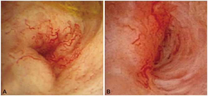
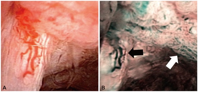
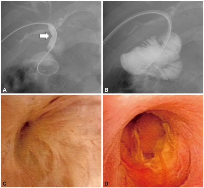
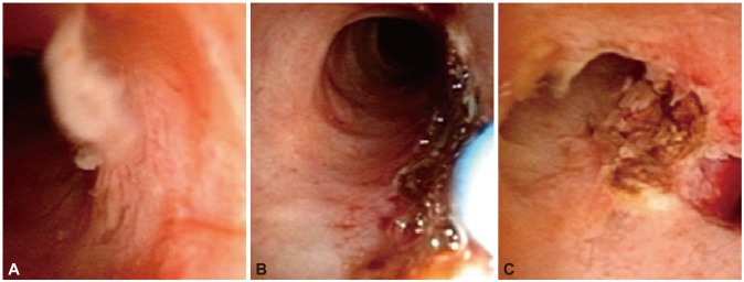
Percutaneous transhepatic cholangioscopy (PTCS) is the most widely used modality for diagnosis and treatment of biliary disease. Although many other novel technologies have been developed based on recent advances in endoscopy, PTCS has its own role. In diagnostics, PTCS is used for evaluation of indeterminate biliary strictures, bile duct tumors, and postoperative biliary strictures that cannot be reached by a peroral approach. In therapeutics, the removal of bile duct stones, dilatation of bile duct strictures including postoperative anastomosis site strictures, and local tumor therapy are indications of PTCS. Especially in a therapeutic role, PTCS has the advantage of maneuverability due to a shorter endoscopic length compared to other cholangioscopic modalities. Hence, PTCS has its own indispensable diagnostic and therapeutic roles.
Keywords: Diagnostic role; Percutaneous transhepatic cholangioscopy; Therapeutic role.
Conflict of interest statement
The authors have no financial conflicts of interest.
Figures
References
-
- McIver MA. An instrument for visualizing the interior of the common duct at operation. Surgery. 1941;9:112–114.
-
- Takada T, Kobayashi S, Yamada A, Uchida Y, Hayashi N. A new technique for the diagnosis and therapy of cholangitic hepatic abscesses; percutaneous transhepatic cholangial drainage (auther's transl) Nihon Shokakibyo Gakkai Zasshi. 1974;71:657–665. - PubMed
-
- Ponchon T, Gagnon P, Berger F, et al. Value of endobiliary brush cytology and biopsies for the diagnosis of malignant bile duct stenosis: results of a prospective study. Gastrointest Endosc. 1995;42:565–572. - PubMed
-
- Macken E, Drijkoningen M, Van Aken E, Van Steenbergen W. Brush cytology of ductal strictures during ERCP. Acta Gastroenterol Belg. 2000;63:254–259. - PubMed
Publication types
LinkOut - more resources
Full Text Sources
Other Literature Sources
