

Duodenal mucosa-associated lymphoid tissue lymphomas: two cases and the evaluation of endoscopic ultrasonography
- PMID: 24143321
- PMCID: PMC3797944
- DOI: 10.5946/ce.2013.46.5.563
Duodenal mucosa-associated lymphoid tissue lymphomas: two cases and the evaluation of endoscopic ultrasonography
Abstract
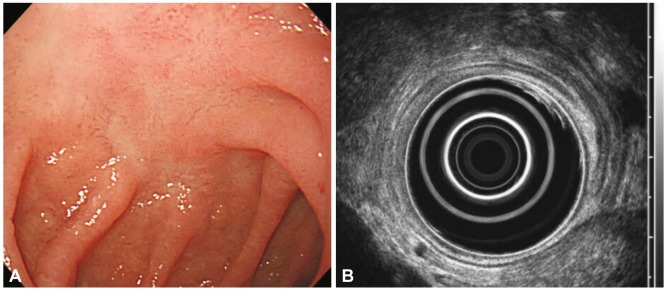
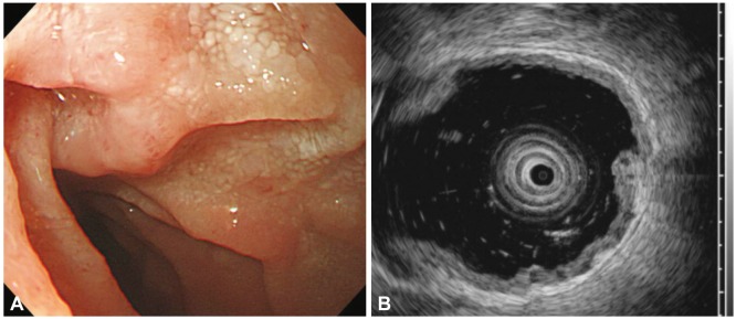
Mucosa-associated lymphoid tissue lymphoma mainly arises in the stomach, with fewer than 30% arising in the small intestine. We describe here two cases of primary duodenal mucosa-associated lymphoid tissue lymphoma which were evaluated by endoscopic ultrasonography. A 52-year-old man underwent endoscopy due to abdominal pain, which demonstrated a depressed lesion on duodenal bulb. Endoscopic ultrasonographic finding was hypoechoic lesion invading the submucosa. The other case was a previously healthy 51-year-old man. Endoscopy showed a whitish granular lesion on duodenum third portion. Endoscopic ultrasonography image was similar to the first case, whereas abdominal computed tomography revealed enlargement of multiple lymph nodes. The first case was treated with eradication of Helicobacter pylori, after which the mucosal change and endoscopic ultrasound finding were normalized in 7 months. The second case was treated with cyclophosphamide, vincristine, prednisolone, and rituximab every 3 weeks. After 6 courses of chemotherapy, the patient achieved complete remission.
Keywords: Duodenum; Endosonography; Lymphoma, B-cell, marginal zone; Mucosa-associated lymphoid tissue.
Conflict of interest statement
The authors have no financial conflicts of interest.
Figures


Similar articles
-
Duodenal mucosa-associated lymphoid tissue lymphoma: a case report.Korean J Intern Med. 2007 Dec;22(4):296-9. doi: 10.3904/kjim.2007.22.4.296. Korean J Intern Med. 2007. PMID: 18309692 Free PMC article.
-
Predictive value of endoscopy and endoscopic ultrasonography for regression of gastric diffuse large B-cell lymphomas after Helicobacter pylori eradication.Dig Endosc. 2009 Oct;21(4):219-27. doi: 10.1111/j.1443-1661.2009.00896.x. Dig Endosc. 2009. PMID: 19961519
-
One patient, two lymphomas. Simultaneous primary gastric marginal zone lymphoma and primary duodenal follicular lymphoma.Arch Pathol Lab Med. 2004 Sep;128(9):1035-8. doi: 10.5858/2004-128-1035-OPTL. Arch Pathol Lab Med. 2004. PMID: 15335250
-
Mucosa-associated lymphoid tissue lymphoma of the duodenum: report of a case resistant to Helicobacter pylori eradication.Hepatogastroenterology. 2004 May-Jun;51(57):732-5. Hepatogastroenterology. 2004. PMID: 15143903 Review.
-
Helicobacter pylori infection in gastric mucosa-associated lymphoid tissue lymphoma.World J Gastroenterol. 2014 Mar 21;20(11):2751-9. doi: 10.3748/wjg.v20.i11.2751. World J Gastroenterol. 2014. PMID: 24659867 Free PMC article. Review.
Cited by
-
Review of lymphoma in the duodenum: An update of diagnosis and management.World J Gastroenterol. 2023 Mar 28;29(12):1852-1862. doi: 10.3748/wjg.v29.i12.1852. World J Gastroenterol. 2023. PMID: 37032723 Free PMC article. Review.
-
Early, Isolated Duodenal Mucosa-Associated Lymphoid Tissue Lymphoma Presenting without Symptoms or Grossly Apparent Endoscopic Lesions and Diagnosed by Random Duodenal Biopsies.Case Rep Gastroenterol. 2016 Jun 27;10(2):323-31. doi: 10.1159/000447293. eCollection 2016 May-Aug. Case Rep Gastroenterol. 2016. PMID: 27482191 Free PMC article.
-
Primary Gastric and Duodenal Mucosa-Associated Lymphoid Tissue Lymphoma With Symptomatic Anemia.ACG Case Rep J. 2024 Jul 17;11(7):e01438. doi: 10.14309/crj.0000000000001438. eCollection 2024 Jul. ACG Case Rep J. 2024. PMID: 39021713 Free PMC article.
References
-
- Isaacson P, Wright DH. Malignant lymphoma of mucosa-associated lymphoid tissue: a distinctive type of B-cell lymphoma. Cancer. 1983;52:1410–1416. - PubMed
-
- Isaacson P, Chott A, Nakamura S, et al. Extranodal marginal zone B-cell lymphoma of mucosa-associated lymphoid tissue (MALT lymph oma) In: Swerdlow SH, Campo E, Harris NL, et al., editors. WHO Classifi cation of Tumours of Haematopoietic and Lymphoid Tissues. Lyon: In ternational Agency for Research on Cancer; 2008. pp. 214–217.
-
- Song IS, Choi KW, Kim CY, et al. Clinicopathologic study of primary gastric lymphoma of B-cell lymphoma of MALT. Korean J Gastroenterol. 1998;31:463–476.
-
- Yokoi T, Nakamura T, Nakamura S. Differential diagnosis of the superficial-type gastric malignant lymphoma: differential diagnosis of gastric MALT lymphomas. Stomach Intest. 2001;36:13–20.
-
- Mehra M, Agarwal B. Endoscopic diagnosis and staging of mucosa-associated lymphoid tissue lymphoma. Curr Opin Gastroenterol. 2008;24:623–626. - PubMed
LinkOut - more resources
Full Text Sources
Other Literature Sources
