

Gastric somatostatinoma: an extremely rare cause of upper gastrointestinal bleeding
- PMID: 24143326
- PMCID: PMC3797949
- DOI: 10.5946/ce.2013.46.5.582
Gastric somatostatinoma: an extremely rare cause of upper gastrointestinal bleeding
Abstract
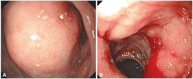
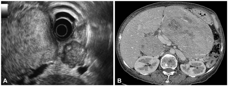
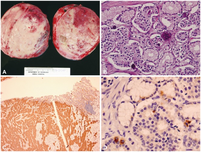
A 49-year-old woman presented with chronic abdominal discomfort, significant weight loss, and chronic intermittent diarrhea. She suddenly developed massive upper gastrointestinal bleeding and was referred for further treatment. Endoscopy indicated a large mass in the upper gastric body with antral and duodenal bulb involvement. Endosonography showed a large well-defined isoechoic gastric subepithelial mass with multiple intra-abdominal and peripancreatic lymphadenopathy, suspected to be malignant on the basis of fine needle aspiration cytology. The tumor was surgically removed, and histopathology showed typical characteristics of a neuroendocrine tumor. On the basis of immunohistochemical staining, somatostatinoma, a rare neuroendocrine tumor, was diagnosed. Gastrointestinal bleeding is a rare presentation and the stomach is an uncommon tumor location.
Keywords: Endosonography; Neuroendocrine tumors; Somatostatinoma; Stomach metastasis.
Conflict of interest statement
The authors have no financial conflicts of interest.
Figures
Similar articles
-
Rare adult gastric duplication cyst mimicking a gastrointestinal stromal tumor.World J Gastroenterol. 2013 Dec 7;19(45):8445-8. doi: 10.3748/wjg.v19.i45.8445. World J Gastroenterol. 2013. PMID: 24363539 Free PMC article.
-
Duodenal somatostatinoma of the ampulla of vater diagnosed by endoscopic fine needle aspiration biopsy: a case report.Acta Cytol. 2001 Jul-Aug;45(4):622-6. doi: 10.1159/000327876. Acta Cytol. 2001. PMID: 11480730
-
Rare Causes of Gastrointestinal Hemorrhage: A Case Series of Adult Duodenal and Jejunal Gastric Heterotopia.Cureus. 2024 Jul 15;16(7):e64604. doi: 10.7759/cureus.64604. eCollection 2024 Jul. Cureus. 2024. PMID: 39144880 Free PMC article.
-
Duodenal somatostatinoma presenting as upper gastrointestinal bleeding.Am J Gastroenterol. 1999 May;94(5):1405-8. doi: 10.1111/j.1572-0241.1999.01097.x. Am J Gastroenterol. 1999. PMID: 10235228 Review.
-
Gastric Inflammatory Fibroid Polyp: A Rare Cause of Occult Upper Gastrointestinal Bleeding.J Investig Med High Impact Case Rep. 2020 Jan-Dec;8:2324709620936840. doi: 10.1177/2324709620936840. J Investig Med High Impact Case Rep. 2020. PMID: 32602395 Free PMC article. Review.
Cited by
-
Somatostatinoma: Beyond neurofibromatosis type 1 (Review).Exp Ther Med. 2020 Oct;20(4):3383-3388. doi: 10.3892/etm.2020.8965. Epub 2020 Jul 3. Exp Ther Med. 2020. PMID: 32905002 Free PMC article. Review.
References
-
- Larsson LI, Hirsch MA, Holst JJ, et al. Pancreatic somatostatinoma: clinical features and physiological implications. Lancet. 1977;1:666–668. - PubMed
-
- Zhang ZY, Zhang R, Wang L, et al. Diagnosis and treatment of pancreatic somatostatinoma: a case report. Chin Med J (Engl) 2008;121:2363–2365. - PubMed
-
- Pernet C, Kluger N, Du-Thanh A, et al. Somatostatin-producing endocrine tumour of the duodenum associated with type 1 neurofibromatosis. Acta Derm Venereol. 2010;90:320–321. - PubMed
-
- Arima H, Natsugoe S, Maemura K, et al. Asymptomatic somatostatinoma of the pancreatic head: report of a case. Surg Today. 2010;40:569–573. - PubMed
-
- Yu RS, Chen Y, Wang LH, Xu XF, Jiang DY. A large functional somatostatinoma in the pancreatic tail: atypical CT appearances. Turk J Gastroenterol. 2009;20:291–294. - PubMed
LinkOut - more resources
Full Text Sources
Other Literature Sources
