. 2013 Dec 15;86(12):1688-98.
doi: 10.1016/j.bcp.2013.10.007. Epub 2013 Oct 19.

Stilbene 5c, a microtubule poison with vascular disrupting properties that induces multiple modes of growth arrest and cell death

Affiliations

Stilbene 5c, a microtubule poison with vascular disrupting properties that induces multiple modes of growth arrest and cell death

M R Alotaibi et al. Biochem Pharmacol. .

Abstract

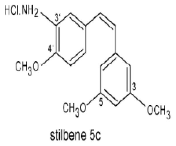
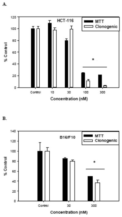
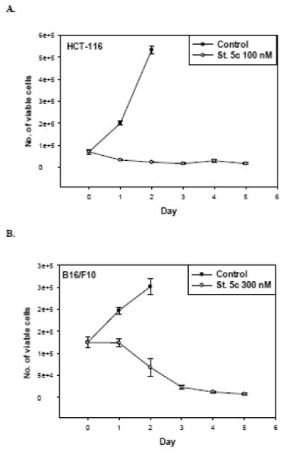
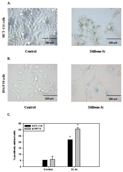
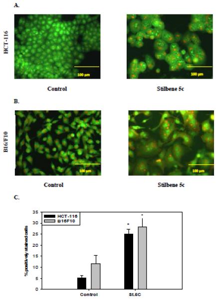
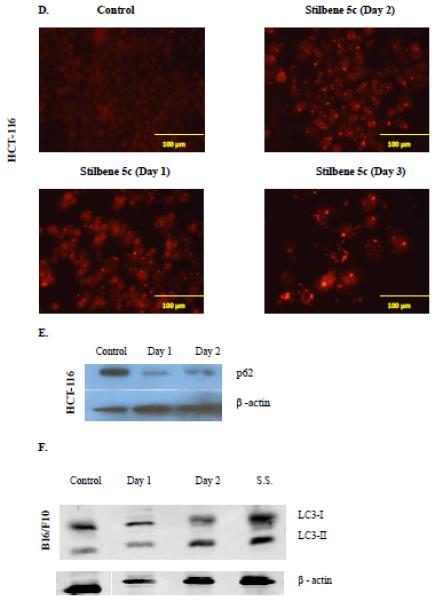
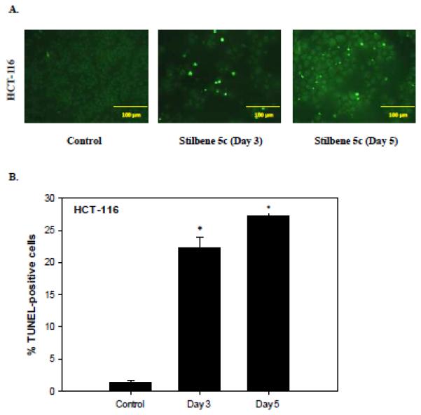
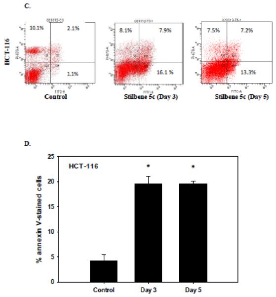
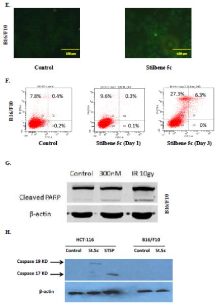
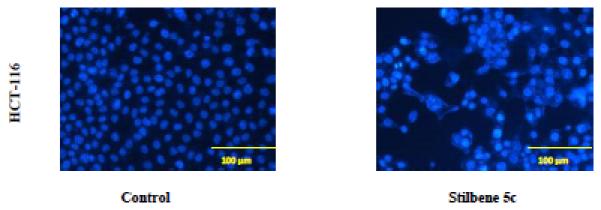
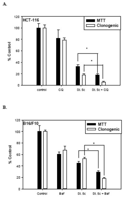
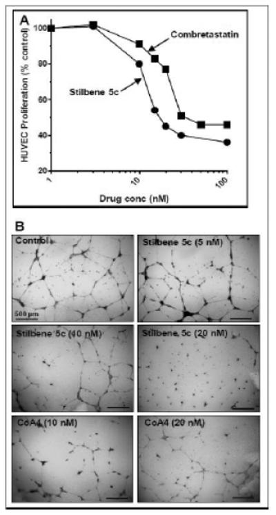
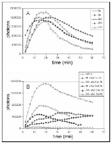
The stilbene derivative, cis-3,4',5-trimethoxy-3'-aminostilbene (stilbene 5c), is a potentially potent antitumor agent that acts via binding to the colchicine-binding site in tubulin. The current studies were designed to investigate the effectiveness of stilbene 5c against the HCT-116 human colon cancer cell line and B16/F10 melanoma cells as well as human endothelial cell tube formation and tumor perfusion. Stilbene 5c produced a time-dependent decrease in cell viability in both cell lines and the capacity of the cells to proliferate was not restored upon removal of the drug. Treatment with stilbene 5c also promoted both senescence and autophagy in both cell lines. TUNEL and annexin 5 staining indicated that apoptosis also occurs in stilbene 5c-treated HCT-116 cells, but not in B16/F10 melanoma cells. DAPI staining revealed morphological changes in the cell nuclei (binucleated and micronucleated cells) indicative of mitotic catastrophe in HCT-116 cells but not in the B16/F10 melanoma cells. p53-null HCT-116 cells demonstrated a similar growth arrest/cell death response to stilbene as p53-wild type HCT-116 cells. Stilbene 5c also completely inhibited human endothelial cell tube formation on Matrigel, consistent with potential anti-angiogenic actions. Using a new method developed for monitoring the pharmacodynamic effects of stilbene 5c in vivo, we found that a single injection of stilbene 5c reduced tumor perfusion by 65% at 4h, returning to baseline by 24h, while subsequent daily injections of stilbene 5c produced progressively larger reductions and smaller rebounds. This work indicates that stilbene 5c could potentially be effective against melanoma and colon cancer through the promotion of multiple modes of growth arrest and cell death coupled with anti-angiogenic and antivascular actions.

Keywords: Angiogenic; Autophagy; Microtubules; Senescence; Vascular disrupting.


Figures

Figure 1
Figure 1
Chemical structure of Stilbene 5c (cis-3, 4', 5-trimethoxy-3'-aminostilbene)
Figure 2
Figure 2. Sensitivity of HCT-116 cells and B16/F10 cells to stilbene 5c
Dose response to stilbene 5c of HCT-116 cells (A) and B16/F10 cells (B) as measured by the MTT assay after 72 hours (closed bars) or clonogenic survival (open bars). Graphs represent pooled data from three replicate experiments. Error bars represent standard error (* p<0.05 compared to control).
Figure 3
Figure 3. Effects of stilbene 5c on growth and survival of HCT-116 cells and B16/F10 melanoma cells
Viability of HCT-116 cells exposed to 100nM stilbene 5c (A) and B16/F10 cells exposed to 300nM stilbene 5c (B) was monitored over a period of 5 days by trypan blue exclusion. (✩) controls (○) Stilbene 5c. 50,000 HCT-116 cells and 100,000 B16F10 cells were plated in each plate of 6-well plates. Graphs represent pooled data from three replicate experiments. Error bars represent standard error (* p<0.05 compared to control).
Figure 4
Figure 4. Senescence in surviving HCT116 colon carcinoma cells and B16/F10 melanoma cells treated with stilbene 5c
HCT-116 cells exposed to 100nM stilbene 5c (A) and B16/F10 cells exposed to 300nM stilbene 5c (B) were incubated with β-galactosidase staining buffer and X-gal. Images shown are representative of three replicate studies. C. Data presented in the graph were generated based on the percentage of cells that were positively stained with β-galactosidase in both treated and non-treated conditions as evaluated by flow cytometry at 72 hours_(* p<0.05 compared to control).
Figure 5
Figure 5. Autophagy induction in HCT116 cells and B16/F10 melanoma cells by stilbene 5c
HCT-116 cells exposed to 100nM stilbene 5c (A) and B16/F10 cells exposed to 300nM stilbene 5c (B) were stained with acridine orange 72 hrs post treatment. All images were taken at 200X and data are representative of three replicate studies. C. The extent of autophagy was determined based on percentage of the cell population showing an increase in acridine orange staining by flow cytometry 72 hrs post treatment (* p<0.05 compared to control). D. Puncta formation in HCT116 cells transfected with an RFP-LC3 vector and treated with 100nM stilbene 5c as shown at 24 hrs, 48 hrs, and 72 hrs. E. Degradation of p62 in HCT-116 cells after exposure to stilbene 5c for 48 hrs. F. Induction of autophagy by western immunoblotting for the microtubule associated light chain protein (LC3-I) conversion to LC3-II in B16/F10 melanoma cells. B16F10 cells were seeded in a serum starved media for 48 hrs and Serum starved cells (S.S.) were used as a positive control.
Figure 6
Figure 6. Assessment of apoptosis in HCT116 colon cancer cells
HCT-116 colon cancer cells were treated with 100nM stilbene 5c. A. TUNEL staining. B. Quantification of TUNEL staining (* p<0.05 compared to control). C. Staining with annexin V and PI . Q1- necrotic cells; Q2- late apoptotic cell population; Q3 – viable cells Q4- early apoptosis. Calculation of the percentage of apoptotic cells presented in Panel D is based on the combined values from Q2 and Q4 (* p<0.05 compared to control). Graphs represent pooled data from three replicate experiments. Error bars represent standard error (* p<0.05 compared to control). (E) Apoptosis evaluated in B16/F10 cells based on the TUNEL assay (F) PI/Annexin staining ( G) PARP cleavage and (H) Caspase 3 activation. For G and H, (IR): radiation (10 Gy) and staurosporine (1 μM) were used as positive control, respectively.
Figure 7
Figure 7. Mitotic catastrophe in HCT116 cells in response to stilbene 5c
DAPI staining of HCT116 colon cancer cells exposed to 100 nM stilbene 5c for 72 hrs. Magnification 200X. Mitotic catastrophe is characterized by bi-nucleated and micro-nucleated cells. In control cells, nuclei are separated and well rounded; treated cell nuclei appear abnormal in size with more than one nucleus in the same proximity. Images are representative of three replicate studies.
Figure 8
Figure 8. Influence of Stilbene 5c on viability of HCT-116 (p53−/−) colon carcinoma cells
HCT-116 (p53−/−) colon cancer cells were treated with 100nM stilbene 5c and viable cell number was determined by trypan blue exclusion. Graphs represent pooled data from three replicate experiments.
Figure 9
Figure 9. Influence of autophagy inhibition on sensitivity to stilbene 5c in HCT-116 cells and B16/F10 melanoma cells
A. Viability and clonogenicity of HCT-116 cells upon exposure to 10 μM chloroquine, 100nM stilbene 5c, and chloroquine + stilbene 5c was measured by the MTT assay and clonogenic survival assay, respectively. B. Viability and clonogenicity of B16/F10 melanoma cells upon exposure to 10 nM bafilomycin, 300nM stilbene 5c, and bafilomycin + stilbene 5c was measured by MTT assay and clonogenic survival assay, respectively. In both assays, cells were pretreated with the autophagy inhibitor for 3 hours prior to stilbene 5c and then exposed to stilbene 5c for 72 hrs (* p<0.05 compared to St.5c alone).
Figure 10
Figure 10. Stilbene 5c inhibits endothelial cell growth and tubule formation
A. HUVEC were plated in 96 well plates and allowed to attach overnight. The indicated concentrations of stilbene 5c (✩) or combretastatin A4 (■) were added, and cell numbers determined after an additional 72 hours by staining with SRB. B. HUVEC were plated on growth-factor reduced Matrigel with the indicated concentrations of stilbene 5c or combretastatin A4. After 24 hours, the wells were fixed, stained and photographed, as described in Methods.
Figure 11
Figure 11. Utilization of luciferase imaging for longitudinal study of tumor perfusion and the effect of stilbene 5c in mouse xenografts
Mice with UCI-101/luciferase tumor xenografts were tested at multiple time points to assess tumor vascular perfusion. A. Luciferin was injected ip at 0, 4, 24, 48, 72 hr and images were collected by Xenogen Live Imaging system every 2 min for total 60 min after injection. In the representative mouse shown, the total number of photons in the tumor area of interest was calculated and plotted against time (min) during the collection period. B. Mice with UCI-101/luciferase tumor xenografts were treated with stilbene 5c at 50 mg/kg/day ip for 3 days. In the mouse shown here, luciferin was injected before, 4 hr after, and 24 hr after the first and second stilbene injections for imaging. Final imaging was done at 24 hr after the third stilbene injection. All images were collected by Xenogen Live Imaging system every 2 min for total 60 min after injection. The total number of photons in the tumor area was calculated and plotted against time (min) during the collection period.

Similar articles

Cited by

References

    1. Ohtsu A. Chemotherapy for metastatic gastric cancer: Past, present, and future. J Gastroenterol. 2008;43:256–64. - PubMed
    1. Tsao H, Atkins MB, Sober AJ. Management of cutaneous melanoma. N Engl J Med. 2004;351:998–1012. - PubMed
    1. Eigentler TK, Caroli UM, Radny P, Garbe C. Palliative therapy of disseminated malignant melanoma: A systematic review of 41 randomised clinical trials. Lancet Oncol. 2003;4:748–59. - PubMed
    1. Avril MF, Aamdal S, Grob JJ, Hauschild A, Mohr P, Bonerandi JJ, et al. Fotemustine compared with dacarbazine in patients with disseminated malignant melanoma: A phase iii study. J Clin Oncol. 2004;22:1118–25. - PubMed
    1. Bedikian AY, Millward M, Pehamberger H, Conry R, Gore M, Trefzer U, et al. Bcl-2 antisense (oblimersen sodium) plus dacarbazine in patients with advanced melanoma: The oblimersen melanoma study group. J Clin Oncol. 2006;24:4738–45. - PubMed

Publication types

Substances