. 2013 Dec;95(3):385-91.
doi: 10.1016/j.yexmp.2013.09.005. Epub 2013 Oct 19.

Extracellular transport of cell-size particles and tumor cells by dendritic cells in culture

Affiliations

Extracellular transport of cell-size particles and tumor cells by dendritic cells in culture

Robert I Thacker et al. Exp Mol Pathol. 2013 Dec.

Abstract

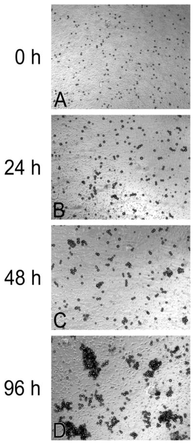
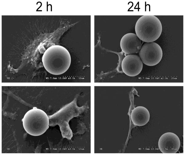
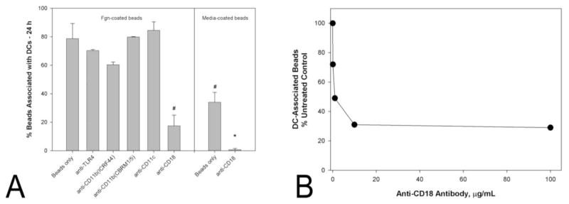
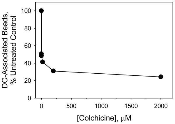
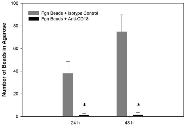
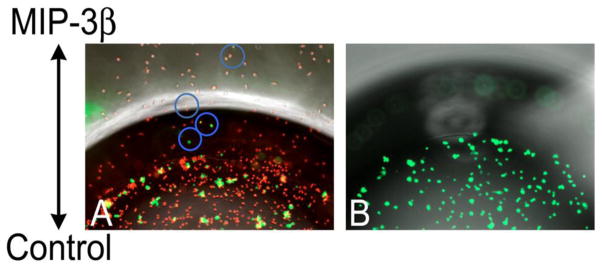
Many particulate materials of sizes approximating that of a cell disseminate after being introduced into the body. While some move about within phagocytic inflammatory cells, others appear to move about outside of, but in contact with, such cells. In this report, we provide unequivocal photomicroscopic evidence that cultured, mature, human dendritic cells can transport in extracellular fashion over significant distances both polymeric beads and tumor cells. At least in the case of polymeric beads, both fibrinogen and the β2-integrin subunit, CD18, appear to play important roles in the transport process. These discoveries may yield insight into a host of disease-related phenomena, including and especially tumor cell invasion and metastasis.

Keywords: Cancer; Cell-size particles; Chemotaxis; Dendritic cells; Directed migration; Extracellular transport; Inflammation; Metastasis; Tumor cells.


Figures

Fig. 1
Fig. 1
Fibrinogen-coated beads, 15.9 ± 2.3 μm, aggregate over time when plated with cultured dendritic cells. Initially, beads co-incubated with dendritic cells are monodisperse, A. Over time, they become progressively more aggregated: B, 24 h; C, 48 h; and D, 96 h.
Fig. 2
Fig. 2
Electron micrographs showing representative extracellular complexes of fibrinogen-coated beads, 15.9 ± 2.3 μm, and dendritic cells, co-incubated for 2 or 24 h.
Fig. 3
Fig. 3
Of the antibodies tested, only one directed against CD18 significantly inhibited complexation of dendritic cells with either fibrinogen-coated beads or media-wetted beads, A. Beads were of diameter 25.7 ± 5.8 μm. Complexation, defined as the association of a bead with 3 or more dendritic cells, was assessed after a 24 h period of co-incubation. Bars indicate the means ± standard deviations of the results of triplicate experiments. The concentration of antibody was in all cases 10 μg/mL. A statistically significant difference, P < 0.001, from fibrinogen-coated beads incubated in the absence of any antibody is indicated by a #. A statistically significant difference, P < 0.001, from media-wetted beads incubated in the absence of any antibody is indicated by a *. Anti-CD18 inhibits complexation of dendritic cells with fibrinogen-coated beads in dose-dependent fashion, B. Beads and dendritic cells were co-incubated in the presence of the antibody for 24 h.
Fig. 4
Fig. 4
Colchicine inhibits complexation of dendritic cells with fibrinogen-coated beads in dose-dependent fashion. Beads and dendritic cells were co-incubated in the presence of the drug for 24 h.
Fig. 5
Fig. 5
In comparison to an isotypic control, antibody directed against CD18 inhibits extracellular transport of fibrinogen-coated beads by dendritic cells. Cells, beads, 15.9 ± 2.3 μm, and antibody, 10 μg/mL, were incubated together within one of two adjacent wells of an agarose field for 2 h prior to the addition of MIP-3β to the other well. Bars indicate the number of beads observed within a high power field located midway between the two wells, 24 and 48 h after the imposition of chemoattractant. Data are the means ± SD’s of the results of 6 separate experiments. * indicates a significant difference between test and control results, P ≤ 0.001. Not shown are data indicating the isotypic control has no influence on bead transport beyond that of medium alone.
Fig. 6
Fig. 6
Fibrinogen-exposed breast tumor cells (green) form aggregates in the presence of dendritic cells (red), A. Over time, 24 h, some tumor cells (within the blue circles) are transported beyond the boundary of the plating well by dendritic cells migrating toward a reservoir of MIP-3β. In the absence of dendritic cells, fibrinogen-exposed tumor cells do not form aggregates, and they do not move toward an imposed chemotactic signal, B.

Similar articles

References

    1. Abbas AK, Lichtman AH. Cellular and Molecular Immunology. Saunders; Philadelphia: 2003a. Antigen Processing and Presentation to T Lymphocytes; pp. 88–90.
    1. Abbas AK, Lichtman AH. Cellular and Molecular Immunology. Saunders; Philadelphia: 2003b. Introduction to Immunology; pp. 24–26.
    1. Altieri DC, et al. Oligospecificity of the cellular adhesion receptor Mac-1 encompasses an inducible recognition specificity for fibrinogen. J Cell Biol. 1988;107:1893–900. - PMC - PubMed
    1. Bachmann MF, et al. Chemokines: more than just road signs. Nat Rev Immunol. 2006;6:159–64. - PubMed
    1. Banchereau J, et al. Immunobiology of dendritic cells. Annu Rev Immunol. 2000;18:767–811. - PubMed

Publication types