. 2013 Oct 28;203(2):205-13.
doi: 10.1083/jcb.201304063. Epub 2013 Oct 21.

Microtubules that form the stationary lattice of muscle fibers are dynamic and nucleated at Golgi elements

Affiliations

Microtubules that form the stationary lattice of muscle fibers are dynamic and nucleated at Golgi elements

Sarah Oddoux et al. J Cell Biol. .

Abstract

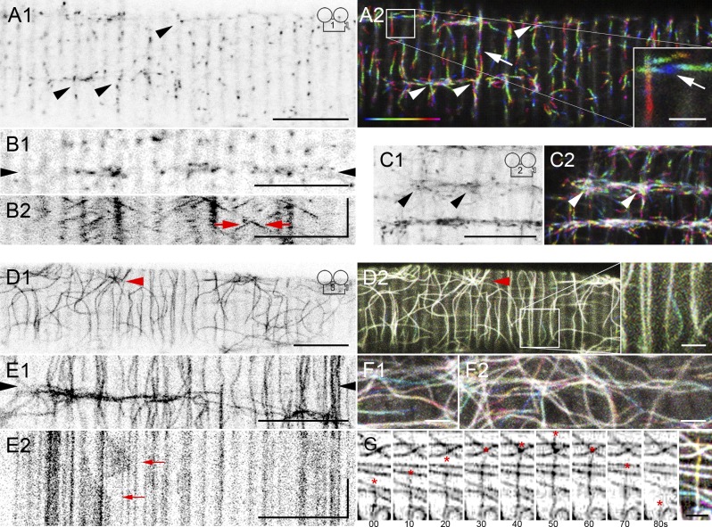
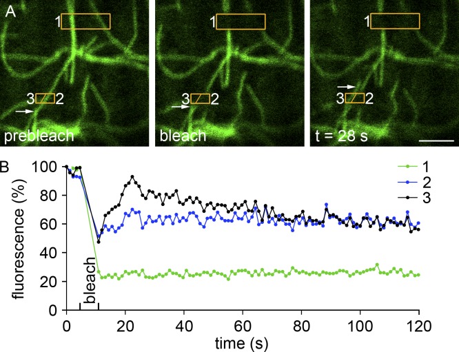
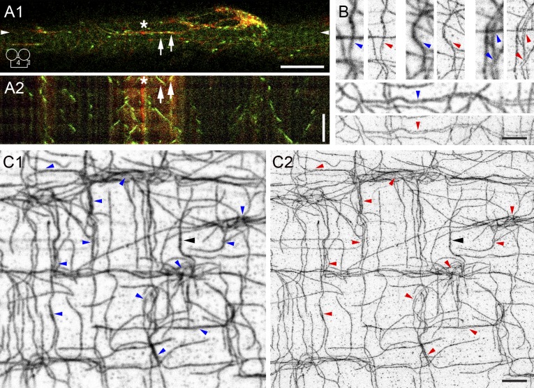
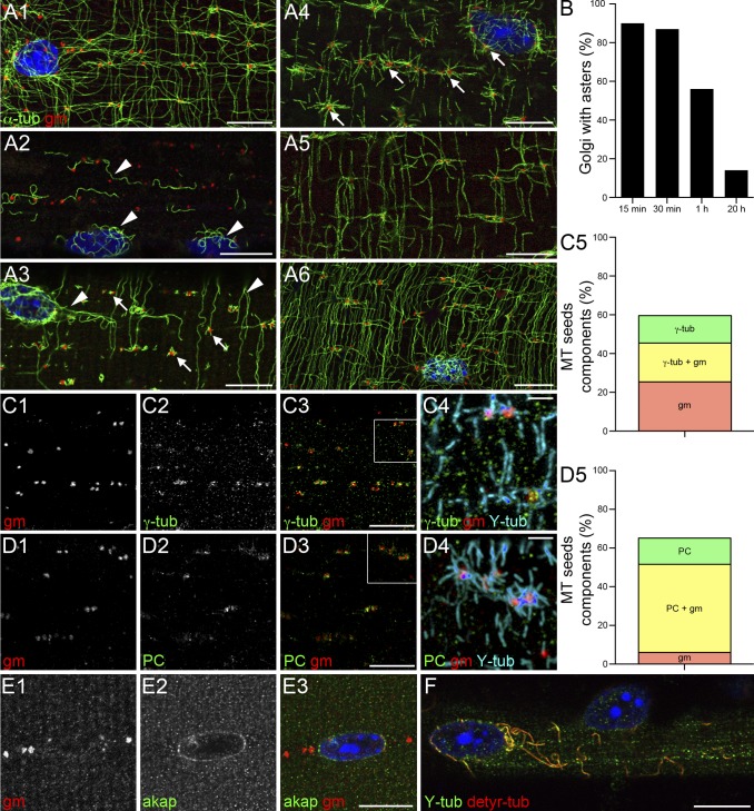
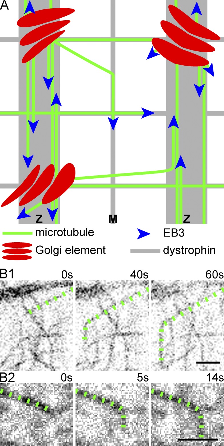
Skeletal muscle microtubules (MTs) form a nonclassic grid-like network, which has so far been documented in static images only. We have now observed and analyzed dynamics of GFP constructs of MT and Golgi markers in single live fibers and in the whole mouse muscle in vivo. Using confocal, intravital, and superresolution microscopy, we find that muscle MTs are dynamic, growing at the typical speed of ∼9 µm/min, and forming small bundles that build a durable network. We also show that static Golgi elements, associated with the MT-organizing center proteins γ-tubulin and pericentrin, are major sites of muscle MT nucleation, in addition to the previously identified sites (i.e., nuclear membranes). These data give us a framework for understanding how muscle MTs organize and how they contribute to the pathology of muscle diseases such as Duchenne muscular dystrophy.


Figures

Figure 1.
Figure 1.
EB3-GFP shows steady MT dynamics ex vivo and in vivo, whereas GFP-tubulin highlights a durable MT frame. A single image, focused near the surface of a plated fiber expressing EB3-GFP (A1), shows typical puncta. The dynamics can be appreciated in the corresponding time-lapse series (Video 1) and in its projection (A2). Color coding of the projection helps to visualize movement: the first image of the series is colored blue and the last one magenta, as in the bar. EB3-GFP puncta mostly move longitudinally and transversely, as if along parallel and antiparallel tracks (A2, arrows). Some MT intersections (arrowheads) seem to behave as MT nucleation sites. An image from another fiber (B1) and the kymograph of the line between the arrowheads (B2; see Materials and methods) indicate that puncta move at the same speed in either direction on the longitudinal axis (oblique lines; red arrows). EB3-GFP dynamics in vivo (C1 and C2; Videos 2 and 5 and Fig. S2) validate plated fibers in all respects (arrowheads point to nucleation sites). In contrast, GFP-tubulin in plated fibers (D–G) appears static: the color-coded projections (D2, F1, and F2), with the same number of images and frame rate as A2, are practically white. An aster (arrowhead) indicates an MT nucleation site. The kymograph (E2) of the line between the arrowheads (E1) shows stationary MTs, with occasional local movement (arrows). Muscle MTs show dynamic instability (G); the asterisk shows the plus end of a MT growing and shrinking over 80 s. See Tables 1 and S1 for data quantitation and technical parameters. Bars: (A–E) 10 µm; (insets) 2 µm; (kymograph vertical time axes) 60 s.
Figure 2.
Figure 2.
FRAP showing only growth (or transport) of MTs restores fluorescence to the bleached area. (A) GFP-tubulin in a plated FDB fiber before (prebleach), just after (bleach), and 28 s after photobleaching of two regions of interest surrounded by orange boxes. For quantitation of recovery (B), the lower box was divided into parts 2 and 3. In seven independent photobleachings, more than half of the bleached boxes recover some fluorescence, as is the case in box 3, by growth or transport of a MT distinct from the original one (A, arrows). Bar, 2 µm.
Figure 3.
Figure 3.
Dynamic MTs grow along MTs and are bundled. EB3-GFP and mCherry-tubulin were coexpressed and simultaneously imaged in plated fibers (A1; Video 4). EB3-GFP (green) moves along static mCherry-tubulin tracks (red; A2 kymograph). The asterisk indicates a nucleation spot and the arrows point to EB3-GFP–labeled MTs growing toward each other. G-STED superresolution microscopy of FDB fibers stained for α-tubulin resolves MTs into two or more components (B and C; panels with blue arrowheads show confocal images; panels with red arrowheads show the corresponding G-STED image at a resolution of 65 nm). A black arrowhead points to a probably unresolved MT. Bars: (A) 10 µm; (B and C) 2.5 µm; (A2, vertical time axis) 60 s.
Figure 4.
Figure 4.
MTs are nucleated on Golgi elements that concentrate γ-tubulin and pericentrin. To investigate MT nucleation, plated FDB fibers were treated with NZ to depolymerize MTs and fixed after different periods of recovery. They were then stained with anti–α-tubulin (α-tub) and anti-GM130 (gm) to label MTs and Golgi elements. In a control fiber (A1), Golgi elements are along MTs, especially at crossings, and around nuclei. After NZ, before recovery (A2), only a few curly MTs remain (arrowheads). These contain both detyrosylated and tyrosylated tubulins (detyr-tub and Y-tub; F), indicating the presence of both stable and dynamic MTs. In the first minute of recovery, NZ-resistant MTs elongate (A3, arrowheads) and MT seeds appear around Golgi elements (A3, arrows). A few minutes later, full asters centered on Golgi elements become prominent (A4, arrows). MTs then progressively reform a network (A5 and A6). During early recovery, ∼90% of Golgi elements are at the center of MT asters; at steady-state, only ∼10% are (B; data are from a single representative experiment out of three; n = 100, from five fibers, for each time point). Fibers at early stages of recovery (as shown in A3) were stained with anti-GM130 and with antibodies against γ-tubulin (γ-tub), pericentrin, and AKAP450. γ-Tubulin and pericentrin are concentrated on Golgi elements of MT seeds (C1–C4 and D1–D4); AKAP450 is only detected around nuclei (E1–E3). Most MT seeds are associated with Golgi elements ± γ-tubulin and some with γ-tubulin alone (C5; Table 2). Similar results are obtained for quantitation with pericentrin (D5; Table 3; two independent experiments, nine fibers, 400 MT seeds for C5, and 322 MT seeds for D5). Bars: (A–F) 10 µm; (C4 and D4) 2 µm.
Figure 5.
Figure 5.
Model of MT organization in skeletal muscle fibers at steady-state. (A) MTs nucleating from Golgi elements grow parallel or antiparallel to existing MTs, thereby forming small bundles, which are guided or restricted by the dystrophin bands positioned along Z lines, M bands, and longitudinal stripes. MTs starting at an oblique angle reorient upon contact with other MTs or dystrophin bands, as observed (B) in GFP-tubulin–expressing fibers. The MT lattice that results is both durable and dynamic. Bars, 2 µm.

References

    1. Applegate K.T., Besson S., Matov A., Bagonis M.H., Jaqaman K., Danuser G. 2011. plusTipTracker: Quantitative image analysis software for the measurement of microtubule dynamics. J. Struct. Biol. 176:168–184 10.1016/j.jsb.2011.07.009 - DOI - PMC - PubMed
    1. Ayalon G., Davis J.Q., Scotland P.B., Bennett V. 2008. An ankyrin-based mechanism for functional organization of dystrophin and dystroglycan. Cell. 135:1189–1200 10.1016/j.cell.2008.10.018 - DOI - PubMed
    1. Ayalon G., Hostettler J.D., Hoffman J., Kizhatil K., Davis J.Q., Bennett V. 2011. Ankyrin-B interactions with spectrin and dynactin-4 are required for dystrophin-based protection of skeletal muscle from exercise injury. J. Biol. Chem. 286:7370–7378 10.1074/jbc.M110.187831 - DOI - PMC - PubMed
    1. Bekoff A., Betz W.J. 1977. Physiological properties of dissociated muscle fibres obtained from innervated and denervated adult rat muscle. J. Physiol. 271:25–40 - PMC - PubMed
    1. Brangwynne C.P., MacKintosh F.C., Kumar S., Geisse N.A., Talbot J., Mahadevan L., Parker K.K., Ingber D.E., Weitz D.A. 2006. Microtubules can bear enhanced compressive loads in living cells because of lateral reinforcement. J. Cell Biol. 173:733–741 10.1083/jcb.200601060 - DOI - PMC - PubMed

Publication types

MeSH terms