. 2013 Nov 5;110(45):18309-14.
doi: 10.1073/pnas.1311406110. Epub 2013 Oct 21.

Molecular mechanism underlying ethanol activation of G-protein-gated inwardly rectifying potassium channels

Affiliations

Molecular mechanism underlying ethanol activation of G-protein-gated inwardly rectifying potassium channels

Karthik Bodhinathan et al. Proc Natl Acad Sci U S A. .

Abstract

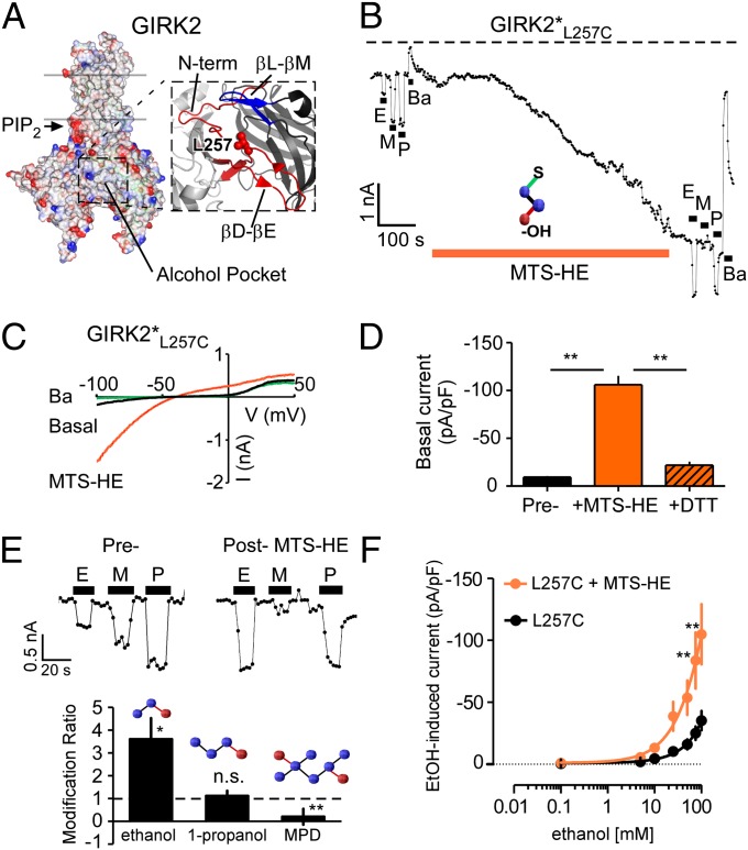
Alcohol (ethanol) produces a wide range of pharmacological effects on the nervous system through its actions on ion channels. The molecular mechanism underlying ethanol modulation of ion channels is poorly understood. Here we used a unique method of alcohol-tagging to demonstrate that alcohol activation of a G-protein-gated inwardly rectifying potassium (GIRK or Kir3) channel is mediated by a defined alcohol pocket through changes in affinity for the membrane phospholipid signaling molecule phosphatidylinositol 4,5-bisphosphate. Surprisingly, hydrophobicity and size, but not the canonical hydroxyl, were important determinants of alcohol-dependent activation. Altering levels of G protein Gβγ subunits, conversely, did not affect alcohol-dependent activation, suggesting a fundamental distinction between receptor and alcohol gating of GIRK channels. The chemical properties of the alcohol pocket revealed here might extend to other alcohol-sensitive proteins, revealing a unique protein microdomain for targeting alcohol-selective therapeutics in the treatment of alcoholism and addiction.

Keywords: Dr-VSP; Kcnj6; Kir3.2; chemical modification; mPhosducin.


Conflict of interest statement

The authors declare no conflict of interest.

Figures

Fig. 1.
Fig. 1.
Alcohol-tagging the pocket constitutively opens GIRK2 channels. (A) Crystal structure (3.6-Å resolution; adapted from ref. 25) of GIRK2 shows L257 (red) in the alcohol pocket, formed by part of N-terminal domain (N-term), βD–βE, and βL–βM loops from two adjacent subunits (blue and red). PIP2 binds at the interface between transmembrane and cytosolic domains (arrow). (B) Plot of inward current through GIRK2*L257C (at −100 mV) shows responses to ethanol (E), MPD (M), 1-propanol (P) (100 mM each), and Ba2+ (1 mM) before and after modification by 1 mM MTS-HE (orange bar). In this and subsequent figures, a dashed line represents zero current level. (C) Current–voltage plots show currents for GIRK2*L257C recorded in extracellular 20K solution (containing 20 mM KCl, 140 mM NaCl, 0.5 mM CaCl2, 2 mM MgCl2, and 10 mM Hepes; pH 7.4, ∼318 mOsm) alone (basal, black), following exposure to 1 mM MTS-HE (orange) and then exposure to Ba2+ (green). (D) Ba2+-sensitive basal GIRK2*L257C current (pA/pF) before and after MTS-HE, and subsequent reversal by DTT (1 mM) (n = 5). **P < 0.01. (E, Upper) Examples of GIRK2*L257C current at −100 mV elicited by alcohols, before and after MTS-HE. (Lower) Bar graph shows MTS-HE modification ratio (Iinduced post/Iinduced pre-MTS) for ethanol (n = 7), 1-propanol (n = 7), and MPD (n = 7). *P < 0.05; **P < 0.01; n.s., not significant (paired Student t test). Dashed line indicates no effect of MTS-HE. (F) Dose–response curves for ethanol-induced current for GIRK2*L257C (pA/pF) before (n = 6; black circles) and after (n = 6; orange circles) MTS-HE. Note the increase in amplitude of current following MTS-HE modification. **P < 0.01 (repeated-measures ANOVA followed by Bonferroni’s post hoc test).
Fig. 2.
Fig. 2.
Chemical diversity rules for activation mediated by the alcohol pocket. (A) Structural view of alcohol pocket in GIRK2 highlighting L257 (red) and S246 (yellow). Cys substitution forms disulfide bond with MTS reagent (MTS-X) carrying ethyl (MTS-E), hydroxyethyl (MTS-HE), benzyl (MTS-F), or hydroxybenzyl (MTS-Y) moieties. (B) Ba2+-sensitive GIRK2*L257C current (pA/pF) before (pre) and after modification by MTS-E (E; 0.1 mM), MTS-HE (HE; 1 mM), MTS-F (F; 0.01 mM), and MTS-Y (Y; 0.1 mM). All MTS reagents except MTS-Y showed significant increase in basal current. **P < 0.01; n.s., not significant. (C) Plot shows inward GIRK2*L257C current (at −100 mV) and responses to 1-propanol (P), MPD (M), ethanol (E) (100 mM each), and 1 mM Ba2+ before and after modification by MTS-F (0.01 mM; light orange bar). (D) Ba2+-sensitive GIRK2*S246c current (pA/pF) before (pre) and after modification with the indicated MTS reagent. All MTS reagents except MTS-F showed no significant change in current. **P < 0.01; n.s., not significant. (E and F) Modification ratio profiles for GIRK2*L257C (E) and GIRK2*S246C (F) for ethanol (Left), 1-propanol (Center), and MPD (Right), after modification by the indicated MTS reagent. *P < 0.05; **P < 0.01; n.s., not significant (paired Student t test).
Fig. 3.
Fig. 3.
MTS-HE activation of GIRK2*L257C channels is independent of Gβγ G proteins. (A) Schematic depicts method for reducing Gβγ (expression of mPhos, +mPhos) or increasing Gβγ (expression of Gβ1γ2, +Gβγ). (B) Plot of inward current through GIRK2*L257C channels (at −100 mV) in HEK293T cells coexpressing mPhos shows alcohol responses and MTS-HE induction. (C) Time course of MTS-HE–dependent activation of GIRK2*L257C currents (normalized current ±SEM vs. time) under reduced (+mPhos; green line), basal (control; blue line), and increased (+Gβγ; red line) levels of Gβγ. (D) Bar graph shows MTS-HE–activated currents for GIRK2*L257C (pA/pF) with +mPhos (n = 5), basal (n = 8), and +Gβγ (n = 7). There was no statistical difference in the amplitude of MTS-HE–activated currents with different levels of Gβγ. n.s., not significant.
Fig. 4.
Fig. 4.
Alcohol tagging inhibits GIRK2*L344C in a G-protein–dependent manner. (A) Structural view of alcohol pocket in GIRK2 highlighting L344 (cyan) in the βL–βM loop, part of the Gβγ binding site (11). (B) Plot of inward current through GIRK2*L344C (at −100 mV) shows inhibition of large basal current with MTS-HE (1 mM). Note the WT-like alcohol activation (rank order: P>M>E) after MTS-HE modification. (C) Current–voltage plots show currents for GIRK2*L344C recorded in extracellular 20K solution alone (basal; black), following exposure to 1 mM MTS-HE (red) and then exposure to 1 mM Ba2+ (green). The large, inwardly rectifying basal GIRK2*L344C current is inhibited by MTS-HE and is also Ba2+-sensitive. (D) Time course of MTS-HE–dependent inhibition of GIRK2*L344C (normalized current ±SEM vs. time) under reduced (+mPhos; n = 7), basal (control; n = 14), and increased (+Gβγ; n = 6) levels of Gβγ. Rate of MTS-HE–dependent inhibition increases with lower Gβγ levels. (E) Ba2+-sensitive GIRK2*L344C current for +mPhos (n = 7), basal (n = 14), and +Gβγ (n = 6) in the absence and then presence of MTS-HE (1 mM). In the unmodified state (–), +mPhos (−40.2 ± 5.3 pA/pF, n = 9) significantly decreased basal GIRK2*L344C currents, compared with control (−109.3 ± 20.1 pA/pF, n = 13) and +Gβγ (−112.1 ± 12.9 pA/pF, n = 6). *P < 0.05; n.s., not significant. The extent of inhibition was indistinguishable for all three conditions (P > 0.05).
Fig. 5.
Fig. 5.
MTS-HE activation of GIRK2*L257C is PIP2-dependent. (A) Schematic shows method for reducing membrane-bound PIP2. Voltage-dependent activation of Dr-VSP (+100 mV) activates the phosphatase, which converts PIP2 to PIP, thus moving GIRK from PIP2-bound open state (green) to unbound closed state (red). (B) Plot of inward current through GIRK2*L257C (at −100 mV) shows MTS-HE–dependent activation and then inhibition following Dr-VSP activation (blue bar). (C) Time course of inhibition for GIRK2*L257C (plot of normalized current vs. time) with variable intervals of Dr-VSP activation (Δt = 5, 20, 30, 50, 100, 300, and 500 ms) before (Left) and after (Right) MTS-HE modification. (D) Plot of time constant (τ) for Dr-VSP–dependent inhibition of GIRK2*L257C vs. depolarization time (longer time corresponds to lower PIP2 levels). Note τ is smaller (indicating faster current decay) for longer depolarization times and is shifted to the right for MTS-HE–modified GIRK2*L257C, suggesting increase in relative PIP2 affinity for the modified channel.
Fig. 6.
Fig. 6.
Alcohol-activated GIRK2 channels exhibit higher relative affinity for PIP2. (A) Plot of inward current through GIRK2* (at −100 mV) shows responses to ethanol (E), MPD (M), 1-propanol (P) (100 mM each), and carbachol (C, 5 μM) before and during activation of Dr-VSP (blue bar). PIP2 depletion decreases both alcohol and carbachol-activated currents. m2 muscarinic receptor was coexpressed with GIRK2*. (B) Bar graph shows amplitude of induced currents for GIRK2* (pA/pF) with ethanol, MPD, 1-propanol (n = 7 each; 100 mM), and carbachol (n = 5; 5 μM) in the absence (−) and then presence (blue bar) of activated Dr-VSP. *P < 0.05; **P < 0.01 (paired Student t test). (C) Plot of inward current through WT GIRK2 channels (at −100 mV) shows transient inhibition with Dr-VSP activation (Δt = 30 ms) in the absence and then presence of 1-propanol (100 mM). A single exponential was fitted to current decay (τ, blue line). (D) Paired measurements of τ for GIRK2 WT in the absence (black circles) and presence (red circles) of 1-propanol (Left) or ethanol (Right). Note that τ is markedly larger in the presence of 1-propanol (n = 12 paired recordings) or ethanol (n = 5 paired recordings), suggesting increase in relative PIP2 affinity; *P < 0.05 (paired Student t test). (E) Control experiment shows small decrease in τ with two consecutive Dr-VSP activation pulses (n = 10 paired recordings). **P < 0.05 (paired Student t test).

Similar articles

Cited by

References

    1. Koob GF, Volkow ND. Neurocircuitry of addiction. Neuropsychopharmacology. 2010;35(1):217–238. - PMC - PubMed
    1. Hyman SE, Malenka RC, Nestler EJ. Neural mechanisms of addiction: The role of reward-related learning and memory. Annu Rev Neurosci. 2006;29:565–598. - PubMed
    1. Aryal P, Dvir H, Choe S, Slesinger PA. A discrete alcohol pocket involved in GIRK channel activation. Nat Neurosci. 2009;12(8):988–995. - PMC - PubMed
    1. Kobayashi T, et al. Ethanol opens G-protein-activated inwardly rectifying K+ channels. Nat Neurosci. 1999;2(12):1091–1097. - PubMed
    1. Lewohl JM, et al. G-protein-coupled inwardly rectifying potassium channels are targets of alcohol action. Nat Neurosci. 1999;2(12):1084–1090. - PubMed

Publication types

MeSH terms

Substances

LinkOut - more resources