. 2013 Nov 5;110(45):18162-7.
doi: 10.1073/pnas.1317049110. Epub 2013 Oct 21.

Hyperammonemia in cirrhosis induces transcriptional regulation of myostatin by an NF-κB-mediated mechanism

Affiliations

Hyperammonemia in cirrhosis induces transcriptional regulation of myostatin by an NF-κB-mediated mechanism

Jia Qiu et al. Proc Natl Acad Sci U S A. .

Abstract

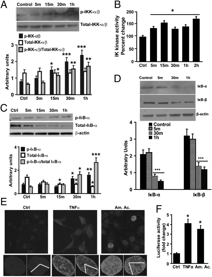
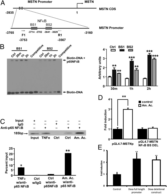
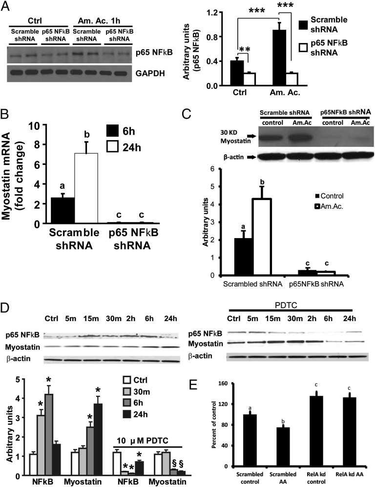
Loss of muscle mass, or sarcopenia, is nearly universal in cirrhosis and adversely affects patient outcome. The underlying cross-talk between the liver and skeletal muscle mediating sarcopenia is not well understood. Hyperammonemia is a consistent abnormality in cirrhosis due to impaired hepatic detoxification to urea. We observed elevated levels of ammonia in both plasma samples and skeletal muscle biopsies from cirrhotic patients compared with healthy controls. Furthermore, skeletal muscle from cirrhotics had increased expression of myostatin, a known inhibitor of skeletal muscle accretion and growth. In vivo studies in mice showed that hyperammonemia reduced muscle mass and strength and increased myostatin expression in wild-type compared with postdevelopmental myostatin knockout mice. We postulated that hyperammonemia is an underlying link between hepatic dysfunction in cirrhosis and skeletal muscle loss. Therefore, murine C2C12 myotubes were treated with ammonium acetate resulting in intracellular concentrations similar to those in cirrhotic muscle. In this system, we demonstrate that hyperammonemia stimulated myostatin expression in a NF-κB-dependent manner. This finding was also observed in primary murine muscle cell cultures. Hyperammonemia triggered activation of IκB kinase, NF-κB nuclear translocation, binding of the NF-κB p65 subunit to specific sites within the myostatin promoter, and stimulation of myostatin gene transcription. Pharmacologic inhibition or gene silencing of NF-κB abolished myostatin up-regulation under conditions of hyperammonemia. Our work provides unique insights into hyperammonemia-induced myostatin expression and suggests a mechanism by which sarcopenia develops in cirrhotic patients.

Keywords: portosystemic shunting; signaling.


Conflict of interest statement

The authors declare no conflict of interest.

Figures

Fig. 1.
Fig. 1.
(A) Skeletal muscle expression of myostatin mRNA showed significantly higher expression in cirrhotics compared with controls (n = 13 each). (B) Representative immunoblots and densitometry of myostatin expression in skeletal muscle from patients with cirrhosis and controls. ***P < 0.001 (cirrhotics compared with controls). Ctrl, control.
Fig. 2.
Fig. 2.
(A and B) Hyperammonemia resulted in reduced muscle mass (A) and grip strength (B) in wild type in contrast to greater muscle mass and increased strength in postdevelopmental myostatin knockout mice. a–b, P < 0.05; a,b–c P < 0.01. (C) Relative fold change in expression of myostatin mRNA in mice in response to hyperammonemia showed significantly increased expression in the myostatin+/+ mice. a–b and a–c, P < 0.001; b–c, P < 0.01. (D) Representative immunoblots for myostatin protein and the densitometry in response to hyperammonemia in myostatin+/+ and myostatin−/− mice. a–b and a–c, P < 0.001; b–c, P < 0.01. n = 5 in each group. AA, ammonium acetate; WT, wild type; KO, postdevelopmental myostatin knockout mice.
Fig. 3.
Fig. 3.
(A) Representative images of phase-contrast microscopy of C2C12 murine myotubes to quantify the myotube diameter. (B) Myotubes treated with 24 h of 10 mM ammonium acetate had significantly lower (P < 0.01) mean diameter (±SEM) compared with control conditions. A total of at least 85 cells in three different plates were quantified. ***P < 0.001 (compared with controls).
Fig. 4.
Fig. 4.
(A) Time course of relative fold change in myostatin mRNA at different concentrations of ammonium acetate in differentiated C2C12 myotubes. Significant increase in myostatin mRNA was observed at all concentrations of ammonium acetate at 6 and 24 h compared with control. (B) Representative immunoblots of the time course of expression of myostatin protein in C2C12 myotubes treated with ammonium acetate (Am. Ac.) and sodium acetate (Na. Ac.) at 10 and 100 mM concentration. C, control. Myostatin expression increased only in response to ammonium acetate. (C) Densitometry of the immunoblots showed increased expression of myostatin protein in a time- and concentration-dependent manner only in response to ammonium acetate, demonstrating that these changes are specific to hyperammonemia. All experiments were performed in triplicate. *P < 0.05; **P < 0.01; ***P < 0.001 (compared with controls).
Fig. 5.
Fig. 5.
(A) Representative immunoblots and densitometry of C2C12 myotubes exposed to 10 mM ammonium acetate and probed for phosphorylation of IKK showed increased phosphorylation of IKK. (B) Time course of percent change in IKK activity in response to 10 mM ammonium acetate in C2C12 myotubes showed significant increase in activity in response to ammonium acetate. (C) Representative immunoblots and densitometry of C2C12 cell myotubes treated with or without (control) 10 mM ammonium acetate for different time points showed increased phosphorylation of IκB-α. (D) Representative immunoblots and densitometry from C2C12 myotubes treated with or without (control) 10 mM ammonium acetate showed degradation of p65 NF-κB binding proteins, IκB-α and -β, during hyperammonemia. (E) Confocal microscopic images of complete and cut sections (yellow V-shaped) of C2C12 myotubes treated with 10 mM ammonium acetate demonstrating nuclear localization of p65 NF-κB (green stain). TNF-α (50 ng/mL) was used as a positive control. All experiments were performed in triplicate. (F) C2C12 myotubes were nuclear transfected with p65 NF-κB −luc-6× (luciferase reporter gene construct controlled by oligomerized κB binding sites). The luciferase fluorescence response was significantly higher in response to both 10 mM ammonium acetate and 50 ng/mL TNF-α (positive control) compared with controls. *P < 0.05; **P < 0.01; ***P < 0.001 (compared with control). All experiments were performed in triplicate.
Fig. 6.
Fig. 6.
(A) Myostatin promoter binding sites for p65 NF-κB and PCR primer design strategy for the analysis of ChIP of myostatin promoter using anti–NF-κB p65. MSTN, myostatin; CDS, coding sequence; BS1, binding site 1; BS2, binding site 2. (B) Representative blots of EMSA and densitometry normalized to controls, demonstrating that p65 NF-κB binds to the myostatin promoter. C2C12 cells were treated with 10 mM ammonium acetate for binding specificity. Arrows indicate the positions of protein–DNA probe complexes and free probes, respectively. C2C12 cells not exposed to ammonium acetate served as negative controls. All experiments were performed in triplicate. BS1, p65 NF-κB binding site 1 on the myostatin promoter; BS2, p65 NF-κB binding site 2 downstream of BS1; Ctrl, control. **P < 0.01; ***P < 0.001 (compared with control). (C) ChIP assay and ChIP real-time PCR demonstrate binding of endogenous p65 NF-κB to the myostatin promoter in C2C12 cells exposed to 10 mM ammonium acetate for 1 h. Cells treated with TNF-α (50 ng/mL) for 30 min served as positive controls. Anti-p65 NF-κB antibody was used for ChIP analysis, and IgG was used as a negative control. As described in Materials and Methods, primers 1 and 2 correspond to a segment covering the two p65 NF-κB binding sequences within 185-bp myostatin promoter. (Upper) Representative gels showing precipitated chromatin. (Lower) Representative quantitative real-time PCR of the precipitated chromatin. *P < 0.05; **P < 0.01 (compared with IgG control group). IgG, normal rabbit IgG; anti–NF-κB p65, rabbit polyclonal IgG anti-p65 NF-κB. All experiments were performed in triplicate. (D) Luciferase reporter assay demonstrates that in response to ammonium acetate, nuclear p65 NF-κB binds to the myostatin promoter, and using a deletion construct with the binding sites deleted, the luciferase response with hyperammonemia was not observed. **P < 0.01 (compared with control). All experiments were performed in triplicate. (E) Dexamethasone (100 μM for 6 h) induced a luciferase response in the deletional construct, demonstrating a functional reporter.
Fig. 7.
Fig. 7.
(A) Representative immunoblots and densitometry of C2C12 myotubes stably transfected with nontargeted sequence (scramble) and knockdown with p65 NF-κB shRNA treated with ammonium acetate for 1 h. Approximately 50% knockdown of p65 NF-κB was achieved at the basal state, and the expression of p65 NF-κB did not increase in the knockdown cells. All experiments were performed four times. **P < 0.01 [compared with nontargeted (scramble) shRNA]; ***P < 0.001 (compared with cells transfected with scrambled shRNA in control medium and cells transfected with p65 NF-κB shRNA treated with ammonium acetate). (B) Relative expression of myostatin mRNA in stable p65 NF-κB knockdown and nontargeted transfected C2C12 myotubes exposed to ammonium acetate for 6 and 24 h. Significant increase in myostatin mRNA in myotubes with nontargeted transfection compared with cells with p65 NF-κB knockdown was found. a–b, P < 0.001; b–c, P < 0.001; a–c, P < 0.01. (C) Representative immunoblots and densitometry for myostatin protein in C2C12 myotubes stably transfected with nontargeted (scramble) plasmid or p65 NF-κB shRNA for knockdown showed significant reduction in myostatin protein expression with knockdown of p65 NF-κB. a–b, P < 0.001; b–c, P < 0.001; a–c, P < 0.01. (D) Representative immunoblots and densitometry of the time course of expression of p65 NF-κB in nuclear lysates and myostatin protein in the cytoplasm of C2C12 murine myotubes exposed to 10 mM ammonium acetate. An increase in p65 NF-κB in the nucleus in response to ammonium acetate precedes the expression of myostatin. Upon blocking p65 NF-κB with 10 μM pyrrolidine dithiocarbamic acid (PDTC; inhibitor of activation of NF-κB by IκB-α stabilization), nuclear p65 NF-κB expression and myostatin expression did not increase with ammonium acetate. All experiments were performed in triplicate. *P < 0.001 (compared to control); §P < 0.01 (compared with control). (E) Myotube diameter in C2C12 myotubes stably transfected with p65 NF-κB or scrambled shRNA, differentiated for 48 h, and exposed to ammonium acetate (10 mM) or control. a–b, P < 0.001; a–c, P < 0.001; b–c, P < 0.001.
Fig. 8.
Fig. 8.
Mechanism of ammonia-induced transcriptional up-regulation of skeletal muscle myostatin.

Similar articles

Cited by

References

    1. Periyalwar P, Dasarathy S. Malnutrition in cirrhosis: Contribution and consequences of sarcopenia on metabolic and clinical responses. Clin Liver Dis. 2012;16(1):95–131. - PMC - PubMed
    1. Hepple RT. Muscle atrophy is not always sarcopenia. J Appl Physiol (1985) 2012;113(4):677–679. - PubMed
    1. Montano-Loza AJ, et al. Muscle wasting is associated with mortality in patients with cirrhosis. Clin Gastroenterol Hepatol. 2012;10(2):166–173. - PubMed
    1. Olde Damink SW, Jalan R, Dejong CH. Interorgan ammonia trafficking in liver disease. Metab Brain Dis. 2009;24(1):169–181. - PubMed
    1. Holecek M, Sprongl L, Tichý M. Effect of hyperammonemia on leucine and protein metabolism in rats. Metabolism. 2000;49(10):1330–1334. - PubMed

Publication types