. 2013 Dec 3;110(49):19679-88.
doi: 10.1073/pnas.1309970110. Epub 2013 Oct 21.

Microenvironmental reprogramming by three-dimensional culture enables dermal papilla cells to induce de novo human hair-follicle growth

Affiliations

Microenvironmental reprogramming by three-dimensional culture enables dermal papilla cells to induce de novo human hair-follicle growth

Claire A Higgins et al. Proc Natl Acad Sci U S A. .

Abstract

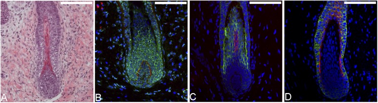
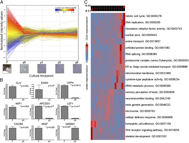
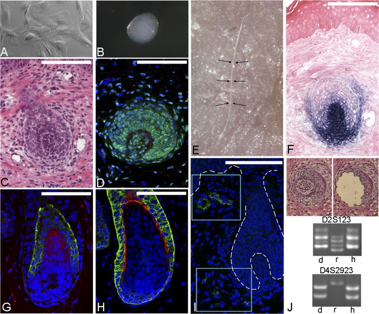
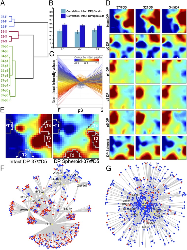
De novo organ regeneration has been observed in several lower organisms, as well as rodents; however, demonstrating these regenerative properties in human cells and tissues has been challenging. In the hair follicle, rodent hair follicle-derived dermal cells can interact with local epithelia and induce de novo hair follicles in a variety of hairless recipient skin sites. However, multiple attempts to recapitulate this process in humans using human dermal papilla cells in human skin have failed, suggesting that human dermal papilla cells lose key inductive properties upon culture. Here, we performed global gene expression analysis of human dermal papilla cells in culture and discovered very rapid and profound molecular signature changes linking their transition from a 3D to a 2D environment with early loss of their hair-inducing capacity. We demonstrate that the intact dermal papilla transcriptional signature can be partially restored by growth of papilla cells in 3D spheroid cultures. This signature change translates to a partial restoration of inductive capability, and we show that human dermal papilla cells, when grown as spheroids, are capable of inducing de novo hair follicles in human skin.


Conflict of interest statement

The authors declare no conflict of interest.

Figures

Fig. 1.
Fig. 1.
Human hair neogenesis instructed by intact dermal papillae. (A) Intact human dermal papillae induced de novo follicle growth in recipient tissue as determined by hematoxylin/eosin staining. (B) A human-specific antibody (green) shows the presence of human cells throughout new hair follicles whereas laminin 5 (red) demarcates the newly formed basement membrane. DAPI (blue) marks all cells. (C) Keratin 71 (green) marks the inner root sheath whereas Keratin 31 (red) marks the hair cortex of new hair follicles. (D) Keratin 14 (green) shows the presence of outer root sheath whereas Keratin 75 (red) marks the companion layer. (Scale bars: 100 µm.)
Fig. 2.
Fig. 2.
Rapid loss of the dermal papilla transcriptome by growth in vitro. (A) Profile plot of dermal papilla culture. Each line represents a transcript that was both significantly and differentially regulated between intact papilla (F), and cells at different time points in culture, from explants (p0), through to passaged cells (p1, p3, p5). (B) Semi-quantitative real-time PCR results showing differences between freshly isolated papillae (F) and papilla cell explants (p0). (C) Global iPAGE analysis showing over- and underrepresented gene ontology terms. Expression bins divide transcripts, with bins on the left containing genes highly expressed within intact papillae and those on the right containing genes up-regulated within cells at p0.
Fig. 3.
Fig. 3.
Human hair follicle neogenesis instructed by papilla spheroids. (A) Dermal papilla cells grown in regular flat culture conditions. (B) A dermal papilla spheroid, established after a 30-h hanging drop culture. (C) Haematoxyin/eosin histology of hair-follicle induction in recipient foreskin tissue. (D) A human-specific antibody (green) shows human cells throughout a de novo follicle whereas laminin 5 (red) demarcates the new basement membrane separating the papilla and matrix, and DAPI (blue) marks all cell nuclei. (E) Macroscopic view of unpigmented hair shaft (between arrows) protruding from experimental foreskin tissue 6 wk post grafting. (F) Alkaline phosphatase staining of the newly formed dermal papilla and sheath. (G) Keratin 71 (green) labels inner root sheath whereas Keratin 31 (red) labels the hair cortex of induced hair follicles. (H) Keratin 14 (green) demarcates the outer root sheath whereas Keratin 75 (red) shows the presence of a companion layer in new hair follicles. (I) Within our human skin grafts, we observed mouse CD31 (green) expression, particularly around the de novo follicles in both the areas near the bulge, and the bulb (boxed regions). Basement membrane delineating hair follicle is outlined with white dashes. (J) Image showing laser-captured tissue and subsequent gel images of microsatellite marker analysis demonstrates that donor spheres share a molecular fingerprint with hair follicles in recipient tissue. d, donor spheres; h, dermal papilla in novel hair induction assay; r, recipient tissue. (Scale bars: 100 µm.)
Fig. 4.
Fig. 4.
Restoration of in vivo signature by reassembly of dermal papilla cells into spheroids. (A) Dendrogram of an unsupervised hierarchical cluster performed on global profiles of intact papillae (blue), cells in culture (green), and dermal spheroids (red). Arrays are identified by patient age. (B) Bar chart comparing similarity of intact papilla with p3 cells, or p3 spheroids, plotted as Pearson’s correlation coefficients. Individual samples are identified by age of tissue donor (37, 32, 34 y). (C) Profile plot comparing intact papillae (F), cells at p3, and spheroids (S) shows that 22% of transcripts differentially regulated between intact papillae and cells at p3 were restored by reassembly into spheroids. (D) GEDI global analysis among microarrays of fresh papillae, cells in culture (p0, p1, p3, p5), and spheroids. Expression values of each probe set, for each sample, were input into a 25 × 26 node grid, whereby changing expression patterns within intact dermal papillae can be assessed as a genome-wide response to growth in culture. (E) GEDI analysis revealed four territories of interest (T1–T4), in which the dynamics of expression changes among samples were of particular interest. (F) Network presentation of transcription factors (yellow nodes) with significant enrichment of genes in territories 1 + 3, or in territories 2 + 4 (G). Red nodes denote territories 1 and 2 in their respective regulons, and blue nodes denote territories 3 and 4. Several transcription factors were detected; however, the transcription factor FLI1 was identified as a regulator unique to the biomarker panel of both territories 1 and 3 and a significantly larger portion of the territory genes than other candidates. In analyzing territories 2 + 4, several significant master regulators were identified with comparable effect sizes, and are indicated within the figure.

Comment in

Similar articles

Cited by

References

    1. Pinkus H. In: The Biology of Hair Growth. Montagna W, Ellis RA, editors. New York: Academic; 1958. pp. 1–32.
    1. Jacobson CM. A comparative study of the mechanisms by which X-irradiation and genetic mutation cause loss of vibrissae in embryo mice. J Embryol Exp Morphol. 1966;16(2):369–379. - PubMed
    1. Kollar EJ. The induction of hair follicles by embryonic dermal papillae. J Invest Dermatol. 1970;55(6):374–378. - PubMed
    1. Dhouailly D. Dermo-epidermal interactions between birds and mammals: Differentiation of cutaneous appendages. J Embryol Exp Morphol. 1973;30(3):587–603. - PubMed
    1. Millar SE. Molecular mechanisms regulating hair follicle development. J Invest Dermatol. 2002;118(2):216–225. - PubMed

Publication types

Associated data