. 2013 Nov 5;110(45):18303-8.
doi: 10.1073/pnas.1314145110. Epub 2013 Oct 21.

Neuroprotective Sirtuin ratio reversed by ApoE4

Affiliations

Neuroprotective Sirtuin ratio reversed by ApoE4

Veena Theendakara et al. Proc Natl Acad Sci U S A. .

Abstract

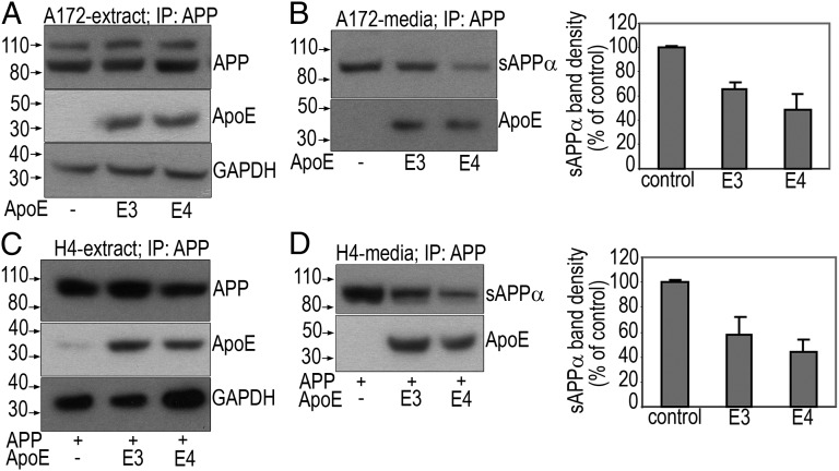
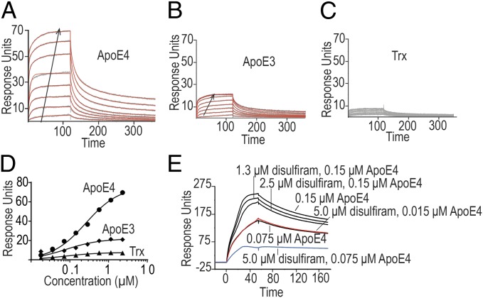
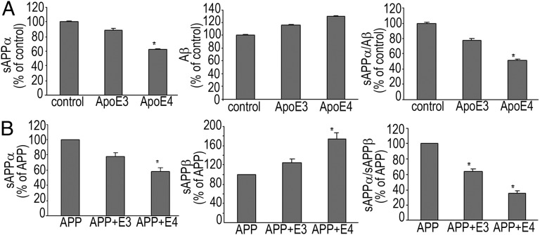
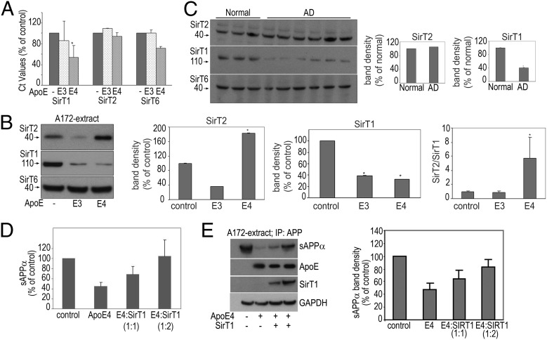
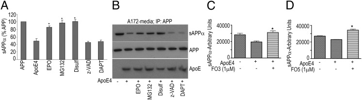
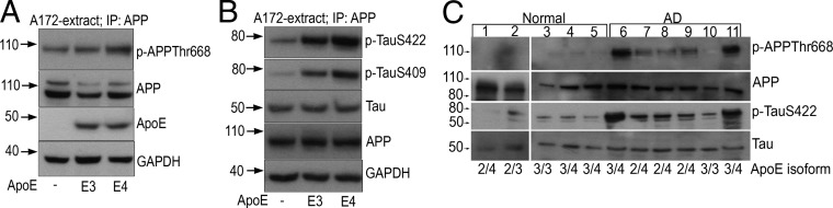
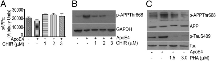
The canonical pathogenesis of Alzheimer's disease links the expression of apolipoprotein E ε4 allele (ApoE) to amyloid precursor protein (APP) processing and Aβ peptide accumulation by a set of mechanisms that is incompletely defined. The development of a simple system that focuses not on a single variable but on multiple factors and pathways would be valuable both for dissecting the underlying mechanisms and for identifying candidate therapeutics. Here we show that, although both ApoE3 and ApoE4 associate with APP with nanomolar affinities, only ApoE4 significantly (i) reduces the ratio of soluble amyloid precursor protein alpha (sAPPα) to Aβ; (ii) reduces Sirtuin T1 (SirT1) expression, resulting in markedly differing ratios of neuroprotective SirT1 to neurotoxic SirT2; (iii) triggers Tau phosphorylation and APP phosphorylation; and (iv) induces programmed cell death. We describe a subset of drug candidates that interferes with the APP-ApoE interaction and returns the parameters noted above to normal. Our data support the hypothesis that neuronal connectivity, as reflected in the ratios of critical mediators such as sAPPα:Aβ, SirT1:SirT2, APP:phosphorylated (p)-APP, and Tau:p-Tau, is programmatically altered by ApoE4 and offer a simple system for the identification of program mediators and therapeutic candidates.


Conflict of interest statement

The authors declare no conflict of interest.

Figures

Fig. 1.
Fig. 1.
ApoE and APP interaction. (A and B) Following transfection of A172 cells with ApoE isoforms alone or H4 cells with APP and ApoE isoforms (C and D), IP was performed with an anti–CT-15-APP antibody (A or C, extract) or N-terminal anti-APP antibody (B or D, media) followed by SDS/PAGE and WB to detect sAPPα, APP, or ApoE. The last panel in A or C represents endogenous GAPDH as a loading control before the pull-down. Band densities of sAPPα (B and D) measured by densitometric quantification of film autoradiograms are expressed as a percentage of sAPPα in control untransfected cells.
Fig. 2.
Fig. 2.
Surface plasmon resonance analysis of the binding of trx-eAPP290–624 to trx-ApoE4 (A), trx-ApoE3 (B), or thioredoxin (trx) (C). The sensograms are shown in gray, and the fits are shown in red. The arrows mark the direction of increasing concentration. The effective KD (KD, eff) was calculated with a single site binding model (D). Competition between disulfiram and ApoE4 for the ectodomain of APP was demonstrated by preincubating trx-ApoE4 with varying concentrations of disulfiram and then analyzing the binding of the mixture to a flow cell treated with biotinylated MBP-eAPP19–624 (E). At saturating concentrations of disulfiram (5 µM, ∼10 times the KD of disulfiram for APP), the binding of ApoE4 is reduced by 50% (Fig. S3).
Fig. 3.
Fig. 3.
ApoE4 but not ApoE3 significantly reduces sAPPα and lowers α/β ratio. A172 cells (A) and H4 cells (B) were transfected with ApoE isoforms alone or APP and ApoE isoforms, respectively. sAPPα and sAPPβ secreted into the medium and Aβ1–42 in the cell extracts were assayed as mentioned in SI Materials and Methods. Data (mean ± SE) are from four experiments performed in triplicate, *P < 0.05.
Fig. 4.
Fig. 4.
ApoE’s effects on Sirtuin expression in cells and AD postmortem tissue. Following transfection of A172 cells with ApoE isoforms, cell pellets were collected and used for RNA isolation and PCR or for SDS/PAGE and WB. (A) The real-time PCR cycling was performed as described in SI Materials and Methods. Data (δCt values expressed as percentage of untransfected control) are from three experiments performed in triplicate, *P < 0.05. (B) Cell extracts were subjected to SDS/PAGE and WB to detect SirT1, T2, and T6. Band densities are expressed as a percentage of untransfected control. Data (mean ± SE) are from three independent experiments,*P < 0.05. (C) Representative immunoblots probed for SirT1, T2, and T6 from homogenates of the temporoparietal region of control subjects and AD patients. Band densities are expressed as a percentage of normal human brains. (D and E) Overexpression of SirT1 reverses ApoE4-mediated reduction in sAPPα. Following transfection of A172 cells with ApoE4 and SirT1 (1:1 and 1:2, respectively), sAPPα secreted into the medium (D) was assayed as mentioned in SI Materials and Methods. Cell extracts were subjected to IP with an N-terminal anti-APP antibody followed by SDS/PAGE and WB to detect sAPPα (E). The last three panels represent ApoE, SirT1, and GAPDH (loading control) before the pull-down. Band densities of sAPPα are expressed as a percentage of sAPPα in control untransfected cells.
Fig. 5.
Fig. 5.
ApoE4-mediated reduction in sAPPα levels is reversed by various inhibitors. A172 cells were transfected with ApoE4 in the presence of epoxomicin (5 μM), MG132 (5 μM), disulfiram (20 μM), z-VAD (40 μM), or DAPT (10 μM). (A and B) Cell culture media were collected and either assayed for sAPPα (A) as described in SI Materials and Methods or subjected to IP with an N-terminal anti-APP antibody followed by SDS/PAGE and WB to detect sAPPα (B). FO3 and FO5 (C and D) reverse ApoE4-mediated reduction in sAPPα levels. Twenty-four hours after transfecting A172 cells with ApoE4, culture medium was changed, and F03 (C) or F05 (D) was added. After an additional 24 h, cell culture media were collected and assayed for sAPPα. Data (mean ± SE) are expressed in arbitrary units of sAPPα released into the media (*P < 0.05).
Fig. 6.
Fig. 6.
ApoE4 triggers APP-Thr668 phosphorylation and Tau phosphorylation. (A and B) Twenty-four hours after transfecting A172 cells with ApoE4, cell extracts were subjected to IP with anti-APP antibody followed by SDS/PAGE and WB to detect p-APP (A), p-TauSer422 (B, Top), p-TauSer409 (B, Middle), or Tau (B, Bottom). The last two panels in A represent ApoE and GAPDH (loading control) or APP and GAPDH (loading control) before the pull-down (B). (C) p-APP and p-Tau in AD. A total of 100 μg each of temporoparietal extracts isolated from normal and AD brains (Table S2) were examined by WB using APP, p-APPThr668, Tau, and p-TauSer422 antibodies. Isoform genotyping of ApoE was performed as described in SI Materials and Methods (Fig. S5).
Fig. 7.
Fig. 7.
Inhibition of GSK-3β or CDK5 decreases p-APP or p-Tau. (A) Culture media from ApoE-transfected A172 cells exposed to the GSK-3β inhibitor CHIR9902 were collected and assayed for sAPPα as described in SI Materials and Methods. sAPPα values are depicted as a percentage of control. Data (mean ± SE) are from three independent experiments performed in triplicate (*P < 0.05). (B and C) Extracts were prepared from ApoE-transfected A172 cells exposed to the GSK-3β inhibitor CHIR9902 (B) or CDK5 kinase inhibitor PHA793887 (C) and subjected to SDS/PAGE and WB to detect p-APP (B) or APP, p-APP, Tau, and p-Tau (C).

References

    1. Bredesen DE, John V. Next generation therapeutics for Alzheimer's disease. EMBO Mol Med. 2013;5(6):795–798. - PMC - PubMed
    1. Mahley RW, Weisgraber KH, Huang Y. Apolipoprotein E4: A causative factor and therapeutic target in neuropathology, including Alzheimer’s disease. Proc Natl Acad Sci USA. 2006;103(15):5644–5651. - PMC - PubMed
    1. Harris FM, et al. Carboxyl-terminal-truncated apolipoprotein E4 causes Alzheimer’s disease-like neurodegeneration and behavioral deficits in transgenic mice. Proc Natl Acad Sci USA. 2003;100(19):10966–10971. - PMC - PubMed
    1. Ye S, et al. Apolipoprotein (apo) E4 enhances amyloid beta peptide production in cultured neuronal cells: ApoE structure as a potential therapeutic target. Proc Natl Acad Sci USA. 2005;102(51):18700–18705. - PMC - PubMed
    1. Verghese PB, et al. ApoE influences amyloid-β (Aβ) clearance despite minimal apoE/Aβ association in physiological conditions. Proc Natl Acad Sci USA. 2013;110(19):E1807–E1816. - PMC - PubMed

Publication types