

GATA3: a multispecific but potentially useful marker in surgical pathology: a systematic analysis of 2500 epithelial and nonepithelial tumors
- PMID: 24145643
- PMCID: PMC3991431
- DOI: 10.1097/PAS.0b013e3182a0218f
GATA3: a multispecific but potentially useful marker in surgical pathology: a systematic analysis of 2500 epithelial and nonepithelial tumors
Abstract
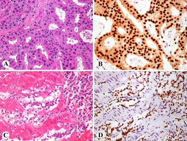
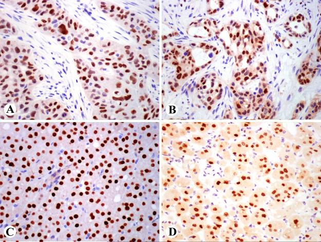
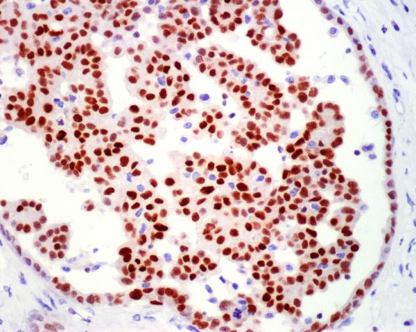
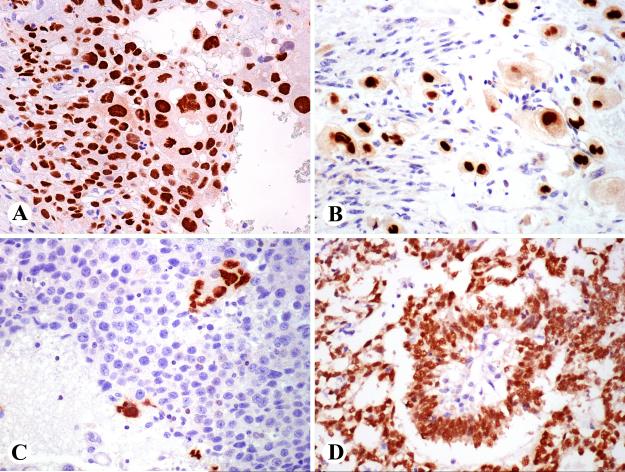
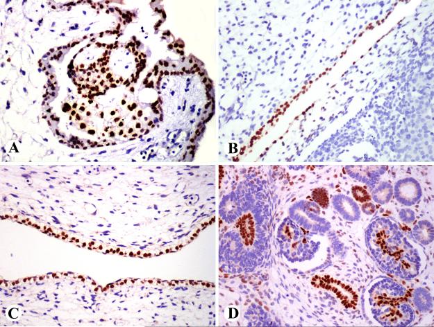
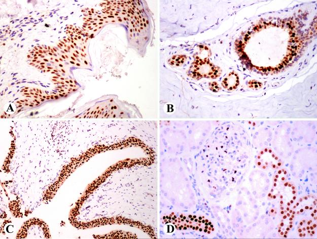
GATA3 is a transcription factor important in the differentiation of breast epithelia, urothelia, and subsets of T lymphocytes. It has been suggested to be useful in the evaluation of carcinomas of mammary or urothelial origin or metastatic carcinomas, but its distribution in normal and neoplastic tissues is incompletely mapped. In this study, we examined normal developing and adult tissues and 2040 epithelial and 460 mesenchymal or neuroectodermal neoplasms for GATA3 expression to explore its diagnostic value in surgical pathology, using monoclonal antibody (clone L50-823) and Leica Bond automated immunohistochemistry. GATA3 was expressed in trophoblast, fetal and adult epidermis, adult mammary and some salivary gland and sweat gland ductal epithelia, urothelia, distal nephron in developing and adult tissues, some prostatic basal cells, and subsets of T lymphocytes. It was expressed stronger in fetal than in adult mesothelia and was absent in respiratory and gastrointestinal epithelia. In epithelial neoplasms, GATA3 was expressed in >90% of primary and metastatic ductal and lobular carcinomas of the breast, urothelial, and cutaneous basal cell carcinomas and trophoblastic and endodermal sinus tumors. In metastatic breast carcinomas, it was more sensitive than GCDFP. Among squamous cell carcinomas, the expression was highest in the skin (81%) and lower in cervical (33%), laryngeal (16%), and pulmonary tumors (12%). Common positivity was found in skin adnexal tumors (100%), mesothelioma (58%), salivary gland (43%), and pancreatic (37%) ductal carcinomas, whereas frequency of expression in adenocarcinomas of lung, stomach, colon, endometrium, ovary, and prostate was <10%. Chromophobe renal cell carcinoma was a unique renal tumor with frequent positivity (51%), whereas oncocytomas were positive in 17% of cases but other types only rarely. Among mesenchymal and neuroectodermal tumors, paragangliomas were usually positive, which sets these tumors apart from epithelial neuroendocrine tumors. Mesenchymal tumors were only sporadically positive, except epithelia of biphasic synovial sarcomas. GATA3 is a useful marker in the characterization of not only mammary and urothelial but also renal and germ cell tumors, mesotheliomas, and paragangliomas. The multiple specificities of GATA3 should be taken into account when using this marker to detect metastatic mammary or urothelial carcinomas.
Figures
References
-
- Asselin-Labat ML, Sutherland KD, Barker H, et al. Gata-3 is an essential regulator of mammary-gland morphogenesis and luminal-cell differentiation. Nat Cell Biol. 2007;9:201–209. - PubMed
MeSH terms
Substances
Grants and funding
LinkOut - more resources
Full Text Sources
Other Literature Sources
