Review
. 2014 Feb;466(2):231-6.
doi: 10.1007/s00424-013-1377-y. Epub 2013 Oct 22.

Post-translational control of cardiac hemodynamics through myosin binding protein C

Affiliations
Review

Post-translational control of cardiac hemodynamics through myosin binding protein C

Manish K Gupta et al. Pflugers Arch. 2014 Feb.

Abstract

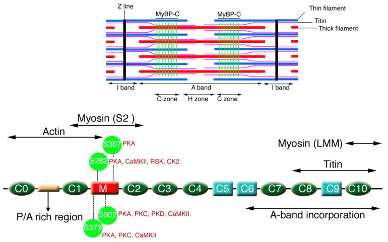
Cardiac myosin binding protein C (cMyBP-C) is an integral sarcomeric protein that associates with the thick, thin, and titin filament systems in the contractile apparatus. Three different isoforms of MyBP-C exist in mammalian muscle: slow skeletal (MyBPC1), fast skeletal (MyBP-C2, with several variants), and cardiac (cMyBP-C). Genetic screening studies show that mutations in MYBPC3 occur frequently and are responsible for as many as 30-35 % of identified cases of familial hypertrophic cardiomyopathy. The function of cMyBP-C is stringently regulated by its post-translational modification. In particular, the addition of phosphate groups occurs with high frequency on certain serine residues that are located in the cardiac-specific regulatory M domain. Phosphorylation of this domain has been extensively studied in vitro and in vivo. Phosphorylation of the M domain can regulate the manner in which actin and myosin interact, affecting the cross bridge cycle and ultimately, cardiac hemodynamics.


Figures

Figure 1
Figure 1
Structure of cMyBP-C. Schematic diagram showing the protein’s restricted sarcomeric location (green lines, top), its domain structure with 8 IgG domains (green), 3 fibronectin domains (light blue) and the cardiac specific M domain (red), with the three phosphorylatable serines discussed in this review indicated. The approximate locations of the binding sites for myosin heavy chain (head and rod regions; S2 and LMM, respectively) are also shown as well as the binding sites for titin and actin.
Figure 2
Figure 2
Schematic description of the effects on cardiac hemodynamics when the three serines are mutated either to non-phosphorylatable alanine or the phosphomimetic aspartate. Heart function was analyzed by invasive catheterization. Detailed data are presented in [13]. Green, yellow and red indicates normal, moderately compromised and poor cardiac function, respectively.

References

    1. Bardswell SC, Cuello F, Rowland AJ, Sadayappan S, Robbins J, Gautel M, Walker JW, Kentish JC, Avkiran M. Distinct sarcomeric substrates are responsible for protein kinase D-mediated regulation of cardiac myofilament Ca2+ sensitivity and cross-bridge cycling. J Biol Chem. 2010;285:5674–5682. doi: 10.1074/jbc.M109.066456. - DOI - PMC - PubMed
    1. Bhuiyan MS, Gulick J, Osinska H, Gupta M, Robbins J. Determination of the critical residues responsible for cardiac myosin binding protein C’s interactions. J Mol Cell Cardiol. 2012;53:838–847. doi: 10.1016/j.yjmcc.2012.08.028. - DOI - PMC - PubMed
    1. Choudhary C, Kumar C, Gnad F, Nielsen ML, Rehman M, Walther TC, Olsen JV, Mann M. Lysine acetylation targets protein complexes and co-regulates major cellular functions. Science. 2009;325:834–840. doi: 10.1126/Science.1175371. - DOI - PubMed
    1. Copeland O, Sadayappan S, Messer AE, Steinen GJ, van der Velden J, Marston SB. Analysis of cardiac myosin binding protein-C phosphorylation in human heart muscle. J Mol Cell Cardiol. 2010;49:1003–1011. doi: 10.1016/j.yjmcc.2010.09.007. - DOI - PubMed
    1. Cuello F, Bardswell SC, Haworth RS, Ehler E, Sadayappan S, Kentish JC, Avkiran M. Novel role for p90 ribosomal S6 kinase in the regulation of cardiac myofilament phosphorylation. J Biol Chem. 2011;286:5300–5310. doi: 10.1074/jbc.M110.202713. - DOI - PMC - PubMed

Publication types

LinkOut - more resources