

Case Reports
doi: 10.1007/s11605-013-2385-0.
Epub 2013 Oct 22.
Surgical management of retroperitoneal leiomyosarcoma arising from the inferior vena cava
Affiliations
- PMID: 24146340
- PMCID: PMC3838601
- DOI: 10.1007/s11605-013-2385-0
Item in Clipboard
Case Reports
Surgical management of retroperitoneal leiomyosarcoma arising from the inferior vena cava
J Gastrointest Surg.
2013 Dec.
Abstract
Retroperitoneal leiomyosarcomas are uncommon tumors, with approximately 300 documented cases in the literature. Management necessitates complete surgical resection in order to offer patients a chance at long-term cure. Resection often presents a challenge as these tumors are often large, involving adjacent structures, and may require reconstruction of the inferior vena cava (IVC). In this article, we will present background information on retroperitoneal leiomyosarcomas and the technical aspects of surgical resection and vascular reconstructive options of the IVC.
Figures
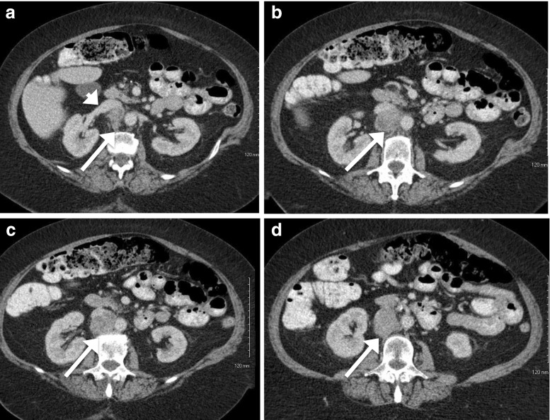
CT images of 4.6 × 4.1 × 4.4 cm leiomyosarcoma. a Image showing the right renal vein entering the IVC (white arrowhead). b, c, d Images showing the extent of IVC invasion (white arrows)
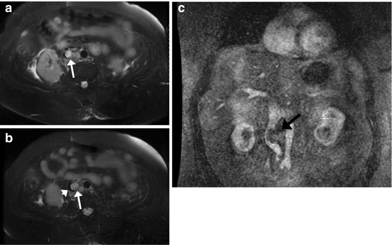
T2-weighted MRI performed after neoadjuvant radiation. a, b A 5.5 cm × 3.3 cm × 3.4 cm infrarenal mass, T2 bright, centered within the posteromedial wall of the inferior vena cava (white arrows) without invasion of the aorta. Note narrowing of IVC lumen (black crescentic lumen, white arrowhead). c Coronal view demonstrating deviation of the IVC (black arrow)
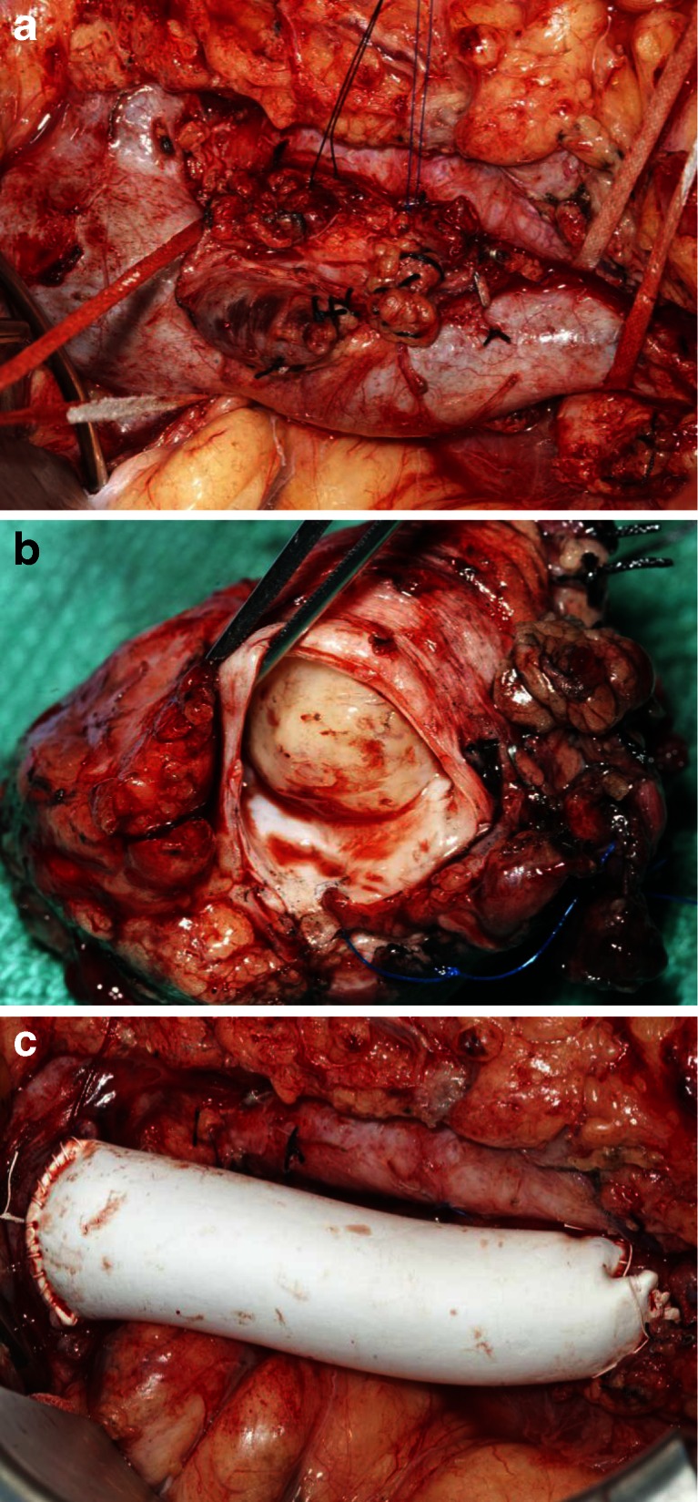
a Intraoperative picture of IVC leiomyosarcoma with the patient's head on the left and the feet on the right. b Resected leiomyosarcoma. c Reconstructed IVC with Gore-Tex graft
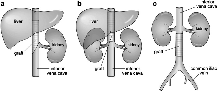
PTFE reconstruction of the IVC. a PTFE reconstruction after right nephrectomy of a level 2 leiomyosarcoma. b PTFE reconstruction of level 2 of the IVC. c PTFE reconstruction of level 3 of the IVC
References
-
- Mingoli A, Cavallaro A, Sapienza P, et al. International registry of inferior vena cava leiomyosarcoma: analysis of a world series on 218 patients. Anticancer Res. 1996;16(5B):3201–5. - PubMed
Publication types
MeSH terms
LinkOut - more resources
Full Text Sources
Other Literature Sources
