

Increase in retinal ganglion cells' susceptibility to elevated intraocular pressure and impairment of their endogenous neuroprotective mechanism by age
- PMID: 24146536
- PMCID: PMC3783363
Increase in retinal ganglion cells' susceptibility to elevated intraocular pressure and impairment of their endogenous neuroprotective mechanism by age
Abstract
Purpose: To investigate age-associated changes in retinal ganglion cell (RGC) response to elevated intraocular pressure (IOP), and to explore the mechanism underlying these changes. Specifically, the effect of aging on inhibitor of apoptosis (IAP) gene family expression was investigated in glaucomatous eyes.
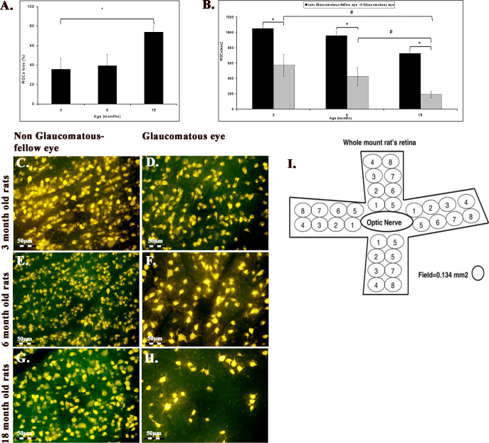
Methods: IOP was induced unilaterally in 82 Wistar rats using the translimbal photocoagulation laser model. IOP was measured using a TonoLab tonometer. RGC survival was evaluated in 3-, 6-, 13-, and 18-month-old animals. Changes in the RNA profiles of young (3-month-old) and old glaucomatous retinas were examined by PCR array for apoptosis; changes in selected genes were validated by real-time PCR; and changes in selected proteins were localized by immunohistochemistry.
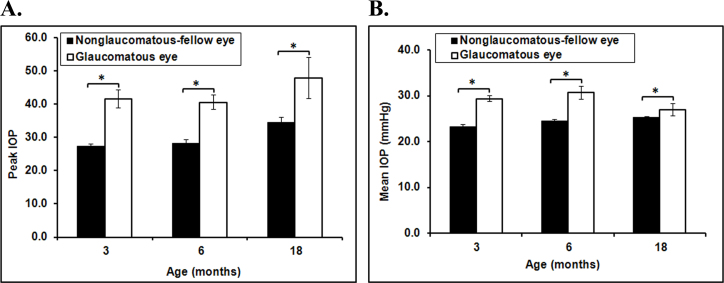
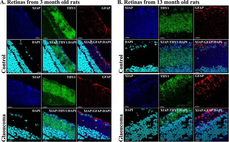
Results: There were no significant IOP differences between the age groups. However, there was a natural significant loss of RGCs with aging and this was more prevalent in glaucomatous eyes. The number of RGCs in glaucomatous eyes decreased from 669±123 RGC/mm² at 3 months to 486±114 RGC/mm² at 6 months and 189±46.5 RGC/mm² at 18 months (n=4-8, p=0.048, analysis of variance). The PCR array revealed different changes in proapoptotic and prosurvival genes between young and old eyes. The two important prosurvival genes, IAP-1 and X-linked IAP (XIAP), acted in opposite directions in 3-month-old and 15-month-old rats, and were significantly decreased in aged glaucomatous retinas, while their expression increased significantly in young glaucomatous eyes. P53 levels did not vary between young glaucomatous and normal fellow eyes, but were reduced with age. B-cell leukemia/lymphoma 2 (Bcl-2) family members and tumor necrosis factor (TNF)-α expression were unaffected by age. Immunohistochemistry results suggested that the sources of changes in IAP-1 protein expression are RGCs and glial cells, and that most XIAP secretion comes from RGCs.
Conclusions: Decreased IAP-1 and XIAP gene expression in aged eyes may predispose RGCs to increased vulnerability to glaucomatous damage. These findings suggest that aging impairs the endogenous neuroprotective mechanism of RGCs evoked by elevated IOP.
Figures


Similar articles
-
Comparison between axonal and retinal ganglion cell gene expression in various optic nerve injuries including glaucoma.Mol Vis. 2013 Dec 16;19:2526-41. eCollection 2013. Mol Vis. 2013. PMID: 24357921 Free PMC article.
-
Global gene expression changes in rat retinal ganglion cells in experimental glaucoma.Invest Ophthalmol Vis Sci. 2010 Aug;51(8):4084-95. doi: 10.1167/iovs.09-4864. Epub 2010 Mar 24. Invest Ophthalmol Vis Sci. 2010. PMID: 20335623 Free PMC article.
-
Experimental glaucoma and optic nerve transection induce simultaneous upregulation of proapoptotic and prosurvival genes.Invest Ophthalmol Vis Sci. 2006 Jun;47(6):2491-7. doi: 10.1167/iovs.05-0996. Invest Ophthalmol Vis Sci. 2006. PMID: 16723461
-
Assessment of retinal ganglion cell damage in glaucomatous optic neuropathy: Axon transport, injury and soma loss.Exp Eye Res. 2015 Dec;141:111-24. doi: 10.1016/j.exer.2015.06.006. Epub 2015 Jun 9. Exp Eye Res. 2015. PMID: 26070986 Review.
-
Glaucoma 2.0: neuroprotection, neuroregeneration, neuroenhancement.Ophthalmology. 2012 May;119(5):979-86. doi: 10.1016/j.ophtha.2011.11.003. Epub 2012 Feb 18. Ophthalmology. 2012. PMID: 22349567 Free PMC article. Review.
Cited by
-
Modeling complex age-related eye disease.Prog Retin Eye Res. 2024 May;100:101247. doi: 10.1016/j.preteyeres.2024.101247. Epub 2024 Feb 15. Prog Retin Eye Res. 2024. PMID: 38365085 Free PMC article. Review.
-
Accelerated Epigenetic Aging Is Associated with Faster Glaucoma Progression: A DNA Methylation Study.Ophthalmology. 2025 May;132(5):550-560. doi: 10.1016/j.ophtha.2024.12.034. Epub 2024 Dec 21. Ophthalmology. 2025. PMID: 39716635
-
Visual field changes after vitrectomy with internal limiting membrane peeling for epiretinal membrane or macular hole in glaucomatous eyes.PLoS One. 2017 May 18;12(5):e0177526. doi: 10.1371/journal.pone.0177526. eCollection 2017. PLoS One. 2017. PMID: 28542230 Free PMC article.
-
Apoptosis in glaucoma: A new direction for the treatment of glaucoma (Review).Mol Med Rep. 2024 May;29(5):82. doi: 10.3892/mmr.2024.13207. Epub 2024 Mar 22. Mol Med Rep. 2024. PMID: 38516770 Free PMC article. Review.
-
The Role of Endogenous Neuroprotective Mechanisms in the Prevention of Retinal Ganglion Cells Degeneration.Front Neurosci. 2018 Nov 15;12:834. doi: 10.3389/fnins.2018.00834. eCollection 2018. Front Neurosci. 2018. PMID: 30524222 Free PMC article. Review.
References
-
- Leung H, Wang JJ, Rochtchina E, Tan AG, Wong TY, Klein R, Hubbard LD, Mitchell P. Relationships between age, blood pressure, and retinal vessel diameters in an older population. Invest Ophthalmol Vis Sci. 2003;44:2900–4. - PubMed
-
- Lin MT, Beal MF. Mitochondrial dysfunction and oxidative stress in neurodegenerative diseases. Nature. 2006;443:787–95. - PubMed
-
- Kong GY, Van Bergen NJ, Trounce IA, Crowston JG. Mitochondrial dysfunction and glaucoma. J Glaucoma. 2009;18:93–100. - PubMed
Publication types
MeSH terms
Substances
LinkOut - more resources
Full Text Sources
Medical
Research Materials
Miscellaneous