Review
. 2013 Oct 17:4:290.
doi: 10.3389/fphys.2013.00290.

Alterations of cAMP-dependent signaling in dystrophic skeletal muscle

Affiliations
Review

Alterations of cAMP-dependent signaling in dystrophic skeletal muscle

Rüdiger Rudolf et al. Front Physiol. .

Abstract

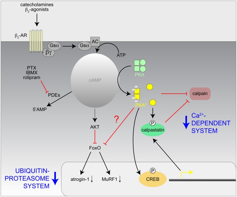
Autonomic regulation processes in striated muscles are largely mediated by cAMP/PKA-signaling. In order to achieve specificity of signaling its spatial-temporal compartmentation plays a critical role. We discuss here how specificity of cAMP/PKA-signaling can be achieved in skeletal muscle by spatio-temporal compartmentation. While a microdomain containing PKA type I in the region of the neuromuscular junction (NMJ) is important for postsynaptic, activity-dependent stabilization of the nicotinic acetylcholine receptor (AChR), PKA type I and II microdomains in the sarcomeric part of skeletal muscle are likely to play different roles, including the regulation of muscle homeostasis. These microdomains are due to specific A-kinase anchoring proteins, like rapsyn and myospryn. Importantly, recent evidence indicates that compartmentation of the cAMP/PKA-dependent signaling pathway and pharmacological activation of cAMP production are aberrant in different skeletal muscles disorders. Thus, we discuss here their potential as targets for palliative treatment of certain forms of dystrophy and myasthenia. Under physiological conditions, the neuropeptide, α-calcitonin-related peptide, as well as catecholamines are the most-mentioned natural triggers for activating cAMP/PKA signaling in skeletal muscle. While the precise domains and functions of these first messengers are still under investigation, agonists of β2-adrenoceptors clearly exhibit anabolic activity under normal conditions and reduce protein degradation during atrophic periods. Past and recent studies suggest direct sympathetic innervation of skeletal muscle fibers. In summary, the organization and roles of cAMP-dependent signaling in skeletal muscle are increasingly understood, revealing crucial functions in processes like nerve-muscle interaction and muscle trophicity.

Keywords: AKAP; PKA; adrenoceptors; dystrophy; endplate; metabolism; neuromuscular junction; skeletal muscle.


Figures

Figure 1
Figure 1
Schematic model of the hypothetic assembly of a PKA microdomain beneath the NMJ. Amongst other potential roles at the NMJ, PKA-RI is critical for proper lifetime regulation of AChR. At least a part of these regulatory processes appears to be linked to the endocytosis/recycling of AChR. From the endocytic compartment, AChRs may either return to the postsynaptic membrane (recycling) or be routed to a degradation pathway. To carry out its function, PKA-RI needs to be recruited to endocytic/recycling vesicles which transport AChR and those vesicles are to be tethered close to the postsynaptic membrane in order to receive local rises of cAMP levels. Anchoring of PKA-RI to these vesicles is mediated by rapsyn, while myo5a serves to restrain endocytic/recycling vesicles in the actin-rich cortex underneath the NMJ. First messengers triggering the relevant local rises in cAMP levels are still elusive and they might originate from motoneurons, sympathetic nervous system or other sites.
Figure 2
Figure 2
PKA-R isoforms display differential distribution patterns in skeletal muscle sarcomeres. Immunohistochemical and GFP-based sensor analyses showed that PKA-RIα essentially co-distributes with microfilaments and PKA-RIIα with m-bands and z-lines. These distribution patterns are the most prevalent in mouse muscle. They are based on AKAP-binding and can be subverted in diseased muscle, such as upon dystrophy. AKAPs relevant for these distribution patterns are still elusive, but myospryn is very likely to participate in the anchoring of PKA-RIIα.
Figure 3
Figure 3
Hypothetical model of the mechanisms involved with the inhibition of the Ca2+-dependent and Ubiquitin-proteasome proteolytic systems in skeletal muscle by catecholamines and β2-agonists. AC, adenylate cyclase; CREB, cAMP response element binding protein; IBMX, isobutylmethylxanthine; PKA*, activated PKA; PTX, pentoxifylline; ?, unknown effect.
Figure 4
Figure 4
Tyrosine hydroxylase (TOH) immunofluorescence is present in sparse axon-like processes and at NMJs. Mouse hindlimb muscles were sectioned and then stained with α-bungarotoxin-AlexaFluor555 (AChR) and an antibody against TOH. Then, confocal microscopy was performed. All panels show maximum z-projections of several optical slices. From left to right, fluorescence signals of AChR, TOH, and overlays are depicted. In overlays, AChR and TOH appear in red and green, respectively. (A) Overview picture showing that most NMJs display enrichments of TOH immunofluorescence. (B) Note thin and pearl chain-like TOH-positive process that ends next to TOH-positive accumulation, which shows a complementary distribution with respect to AChR. (C) Detail of a NMJ with TOH staining complementary to AChR labelling and with emanating axon-like process.
Figure 5
Figure 5
β2-AR-immunofluorescence is found in motoneurons and muscle fibers and is severely altered in dystrophic muscle. Mouse hindlimb muscles of wildtype (A,B,E left) or dystrophic mdx mice (C,D,E right) were sectioned and then stained with α-bungarotoxin-AlexaFluor555 (AChR) and an antibody against β2-AR. Then, confocal microscopy was performed. (A–D) Show maximum z-projections of several optical slices, in (E) single optical slices are depicted. From left to right, fluorescence signals of AChR, β2-AR, and overlays are depicted. In overlays, AChR and β2-AR appear in red and green, respectively. In wildtype muscles, β2-AR immunofluorescence covers entire motor nerve bundles (A) and perfectly matches the AChR arborized structures in the NMJ (B). This is typical for the distribution of motoneuronal markers. Conversely, β2-AR immunofluorescence is much sparser in dystrophic muscle (C) and exhibits only partial overlap with AChR staining (D). In muscle fibers of wildtype animals (E left) β2-AR is found in triple striations per sarcomer, similar to the distribution of PKA-RIIα (see Figure 2). This striation is mostly absent in dystrophic muscle (E right), where β2-AR distribution is often uniform along the fibers. Finally, anostomotic β2-AR-positive, axon-like processes of unknown identity are also often seen running along muscle fibers (E left).

References

    1. Adachi S., Oka J., Nagao T., Fukuda H. (1992). Activation of beta-adrenergic receptor induces Na(+)-dependent inward currents in acutely dissociated motoneurons of bullfrog spinal cord. Brain Res. 571, 79–88 10.1016/0006-8993(92)90511-7 - DOI - PubMed
    1. Akaaboune M., Culican S. M., Turney S. G., Lichtman J. W. (1999). Rapid and reversible effects of activity on acetylcholine receptor density at the neuromuscular junction in vivo. Science 286, 503–507 10.1126/science.286.5439.503 - DOI - PubMed
    1. Altarejos J. Y., Montminy M. (2011). CREB and the CRTC co-activators: sensors for hormonal and metabolic signals. Nat. Rev. Mol. Cell Biol. 12, 141–151 10.1038/nrm3072 - DOI - PMC - PubMed
    1. Apel E. D., Glass D. J., Moscoso L. M., Yancopoulos G. D., Sanes J. R. (1997). Rapsyn is required for MuSK signaling and recruits synaptic components to a MuSK-containing scaffold. Neuron 18, 623–635 10.1016/S0896-6273(00)80303-7 - DOI - PubMed
    1. Arreola J., Calvo J., Garcia M. C., Sanchez J. A. (1987). Modulation of calcium channels of twitch skeletal muscle fibres of the frog by adrenaline and cyclic adenosine monophosphate. J. Physiol. 393, 307–330 - PMC - PubMed

LinkOut - more resources