. 1985;317(6040):809-11.
doi: 10.1038/317809a0.

Voltage-dependent calcium and potassium channels in retinal glial cells

Voltage-dependent calcium and potassium channels in retinal glial cells

E A Newman. Nature. 1985.

Abstract

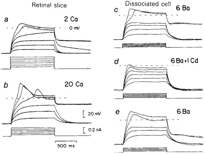
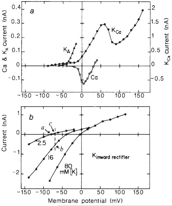
Glial cells, which outnumber neurones in the central nervous system, have traditionally been considered to be electrically inexcitable and to play only a passive role in the electrical activity of the brain. Recent reports have demonstrated, however, that certain glial cells, when maintained in primary culture, possess voltage-dependent ion channels. It remains to be demonstrated whether these channels are also present in glial cells in vivo. I show here that Müller cells, the principal glial cells of the vertebrate retina, can generate 'Ca2+ spikes' in freshly excised slices of retinal tissue. In addition, voltage-clamp studies of enzymatically dissociated Müller cells demonstrate the presence of four types of voltage-dependent ion channels: a Ca2+ channel, a Ca2+-activated K+ channel, a fast-inactivating (type A) K+ channel and an inward-rectifying K+ channel. Currents generated by these voltage-dependent channels may enhance the ability of Müller cells to regulate extracellular K+ levels in the retina and may be involved in the generation of the electroretinogram.


Figures

Fig. 1
Fig. 1
Regenerative ‘Ca2+ spikes’ evoked by depolarizing current pulses in Müller cells of the salamander. a, b, Voltage recordings from a cell in a retinal slice. The regenerative response in control perfusate (a) increased significantly when [Ca2+] was raised from 2 to 20 mM (b). The plateau of the action potential lasted for several seconds following stimulation. The cell was pressure-injected with Cs+ and TEA to reduce the resting K+ conductance and bathed in pH 6.8 perfusate to de-couple the cell from other Müller cells. c-e, Recordings from a dissociated Müller cell in 6 mM Ba2+. Current pulses evoked a large sustained Ba2+ spike (c) which was substantially reduced by addition of 1 mM Cd2+ (d). The regenerative response returned when the Cd2+ was washed out (e). In both cells the resting potential was maintained at -70 mV by injection of small continuous currents. Current pulse amplitudes are indicated below each series of traces.
Fig. 2
Fig. 2
Voltage-clamp records of voltage-dependent currents in dissociated salamander Müller cells. a, Inward Ca2+ current. The cell potential was stepped to the value which evoked maximal inward current (+15 mV in 20 mM Ba2+ and +5 mV in mM Ba2+). The inward current increased when [Ba2+] was raied from 2 to 20 mM and was abolished by addition of 2 mM Cd2+. The perfusate contained 5 mM TEA to reduce Ca2+-activated K+ currents. Recordings made from a cell with endfoot intact. b, Outward Ca2+-activated K+ current. In 10 mM Ca2+ perfusate (10 Ca), a slowly developing outward current was superimposed on the inward Ca2+ current. 1 mM TEA reduced the outward current while 2 mM Cd2+ eliminated both the inward and outward currents. Recordings from a cell lacking its endfoot process. c, Transient outward (type A) K+ current. The transient current was eliminated when the voltage step was preceded by a 2-s prepulse to -40 mV. d, Transient current shown in isolation by subtracting records preceded by a prepulse from records without a prepulse. The ‘control’ is the difference of the two records in c. The transient current in control perfusate was unaltered by 5 mM TEA but abolished by 5 mM 4-AP. Records from c and d were from the same endfoot-shorn Müller cell. e, Inward-rectifying K+ current evoked by depolarizing and hyperpolarizing voltage steps in 80 mM K+ perfusate. Holding potential (Eh), -4 mV, the membrane potential of the cell in 80 mM K+. Recordings from an endfoot-shorn cell. In a-e, inward currents are shown as downward deflections. Capacitive transients were reduced using transient cancellation circuitry. In a and b, linear contributions of the ‘leakage current’ were removed by subtracting a scaled version of records evoked by depolarizations to ∼ -60 mV.
Fig. 3
Fig. 3
Current-voltage plots of the voltage-dependent ionic currents illustrated in Fig. 2. a, The Ca2+ current relation (Ca) was obtained from an intact Müller cell bathed in perfusate containing 2 mM Ba2+ and 5 mM TEA. It represents peak inward current, corrected for an extrapolated linear leakage current. Eh, -80 mV. The Ca2+-activated K+ current relation (KCa) was obtained from an endfoot-shorn cell in control perfusate and represents the steady-state current attained during a 3.5-s voltage pulse. The small outward current on the KCa curve between -80 and -30 mV is inward rectifier current. Eh, -90 mV. The transient (type A) K+ current relation (KA) was obtained from an endfoot-shorn Müller cell in control perfusate. Current amplitude was measured between the peak of the response and the value at the end of an 800 ms pulse. Eh, -90 mV. b, The inward rectifier K+ current relation was recorded from an endfoot-shorn cell bathed sequentially in 2.5, 16 and 80 mM K+ perfusate. Current amplitudes represent the maximal values attained during an 800-ms voltage step. Eh, -80, -40 and -4 mV, the cell membrane potential in the three perfusates. The dashed line and points a-c in b are used to illustrate changes in membrane conductance caused by a localized [K+]o increase. See text for details. Note differences in the vertical scales for the four currents.

References

    1. Kuffler SW. Proc. R. Soc. 1967;B168:1–21. - PubMed
    1. Chiu SY, Schrager P, Ritchie JM. Nature. 1984;311:156–157. - PubMed
    1. MacVicar BA. Science. 1984;226:1345–1347. - PubMed
    1. Bevan S, Raff M. Nature. 1985;315:229–232. - PubMed
    1. Newman EA. Nature. 1984;309:155–157. - PMC - PubMed

Publication types