

Association of pro-inflammatory cytokines and iron regulatory protein 2 (IRP2) with Leishmania burden in canine visceral leishmaniasis
- PMID: 24146743
- PMCID: PMC3795717
- DOI: 10.1371/journal.pone.0073873
Association of pro-inflammatory cytokines and iron regulatory protein 2 (IRP2) with Leishmania burden in canine visceral leishmaniasis
Abstract
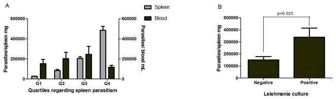
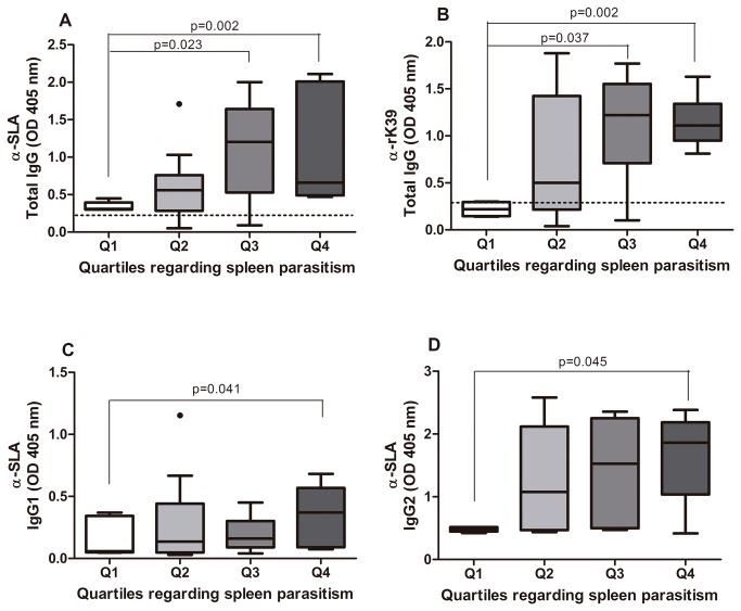
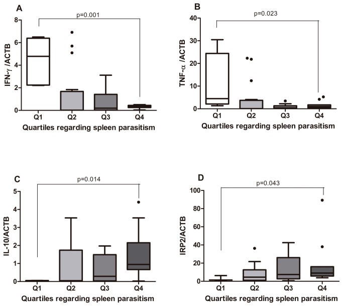
Leishmania infantum infection in humans and dogs can evolve with a wide range of clinical presentations, varying from asymptomatic infections to visceral leishmaniasis. We hypothesized that the immune response elicited by L. infantum infection could modulate whether the host will remain asymptomatic or progress to disease. A total of 44 dogs naturally infected with L. infantum were studied. Leishmania burden was estimated in the blood and spleen by qPCR. The expression of IFN-γ, TNF-α, IL-10 and Iron Regulatory Protein 2 (IRP2) were determined in the spleen by quantitative PCR. Sera cytokines were evaluated by ELISA. Dogs were grouped in quartiles according parasite burden. Increased expression of IFN-γ and TNF-α was associated with reduced Leishmania burden, whereas increased IL-10 and IRP2 expressions were associated with higher Leishmania load. Increased plasma albumin and IFN-γ expression explained 22.8% of the decrease in parasite burden in the spleen. These data confirm that lower IFN-γ response and higher IL-10 correlated with increased parasite load and severity of the visceral leishmaniasis in dogs. The balance between the branches of immune response and the intracellular iron availability could determine, in part, the course of Leishmania infection.
Conflict of interest statement
Figures


Similar articles
-
Evaluation of TNF-α, IL-4, and IL-10 and parasite density in spleen and liver of L. (L.) chagasi naturally infected dogs.Ann Trop Med Parasitol. 2011 Jul;105(5):373-83. doi: 10.1179/1364859411Y.0000000027. Ann Trop Med Parasitol. 2011. PMID: 21929879 Free PMC article.
-
Parasite load induces progressive spleen architecture breakage and impairs cytokine mRNA expression in Leishmania infantum-naturally infected dogs.PLoS One. 2015 Apr 13;10(4):e0123009. doi: 10.1371/journal.pone.0123009. eCollection 2015. PLoS One. 2015. PMID: 25875101 Free PMC article.
-
Expression of regulatory T cells in jejunum, colon, and cervical and mesenteric lymph nodes of dogs naturally infected with Leishmania infantum.Infect Immun. 2014 Sep;82(9):3704-12. doi: 10.1128/IAI.01862-14. Epub 2014 Jun 16. Infect Immun. 2014. PMID: 24935975 Free PMC article.
-
Systemic and compartmentalized immune response in canine visceral leishmaniasis.Vet Immunol Immunopathol. 2009 Mar 15;128(1-3):87-95. doi: 10.1016/j.vetimm.2008.10.307. Epub 2008 Oct 17. Vet Immunol Immunopathol. 2009. PMID: 19054576 Review.
-
Cutaneous immune mechanisms in canine leishmaniosis due to Leishmania infantum.Vet Immunol Immunopathol. 2015 Feb 15;163(3-4):94-102. doi: 10.1016/j.vetimm.2014.11.011. Epub 2014 Nov 20. Vet Immunol Immunopathol. 2015. PMID: 25555497 Review.
Cited by
-
Leishmania infantum-specific production of IFN-γ and IL-10 in stimulated blood from dogs with clinical leishmaniosis.Parasit Vectors. 2016 Jun 3;9(1):317. doi: 10.1186/s13071-016-1598-y. Parasit Vectors. 2016. PMID: 27260142 Free PMC article.
-
Vitamins A and D and Zinc Affect the Leshmanicidal Activity of Canine Spleen Leukocytes.Animals (Basel). 2021 Aug 31;11(9):2556. doi: 10.3390/ani11092556. Animals (Basel). 2021. PMID: 34573521 Free PMC article.
-
Therapeutic control of leishmaniasis by inhibitors of the mammalian target of rapamycin.PLoS Negl Trop Dis. 2018 Aug 22;12(8):e0006701. doi: 10.1371/journal.pntd.0006701. eCollection 2018 Aug. PLoS Negl Trop Dis. 2018. PMID: 30133440 Free PMC article.
-
Humoral Responses and Ex Vivo IFN-γ Production after Canine Whole Blood Stimulation with Leishmania infantum Antigen or KMP11 Recombinant Protein.Vet Sci. 2022 Mar 4;9(3):116. doi: 10.3390/vetsci9030116. Vet Sci. 2022. PMID: 35324844 Free PMC article.
-
Canine Cytokines Profile in an Endemic Region of L. infantum: Related Factors.Vet Sci. 2022 Jun 20;9(6):305. doi: 10.3390/vetsci9060305. Vet Sci. 2022. PMID: 35737357 Free PMC article.
References
Publication types
MeSH terms
Substances
Grants and funding
LinkOut - more resources
Full Text Sources
Other Literature Sources
