

The AERO system: a 3D-like approach for recording gene expression patterns in the whole mouse embryo
- PMID: 24146773
- PMCID: PMC3797748
- DOI: 10.1371/journal.pone.0075754
The AERO system: a 3D-like approach for recording gene expression patterns in the whole mouse embryo
Abstract
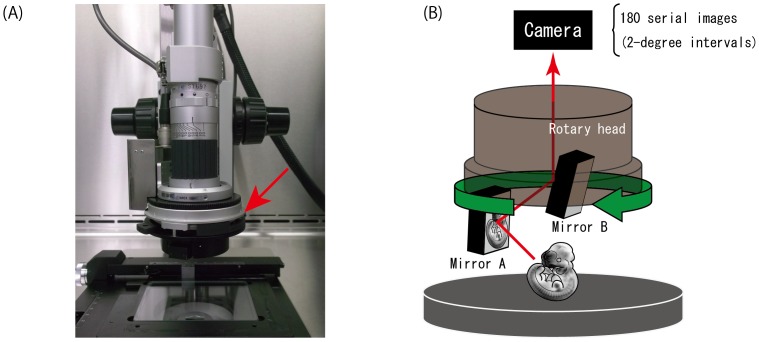
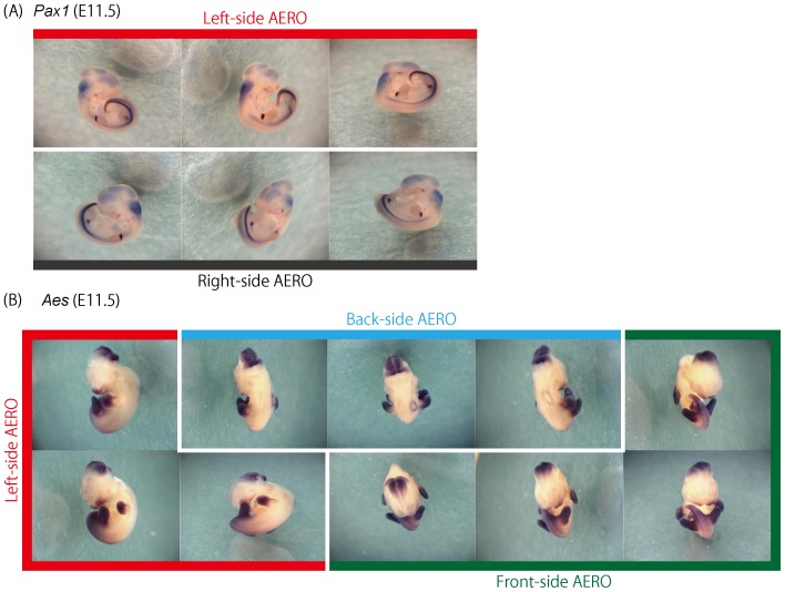
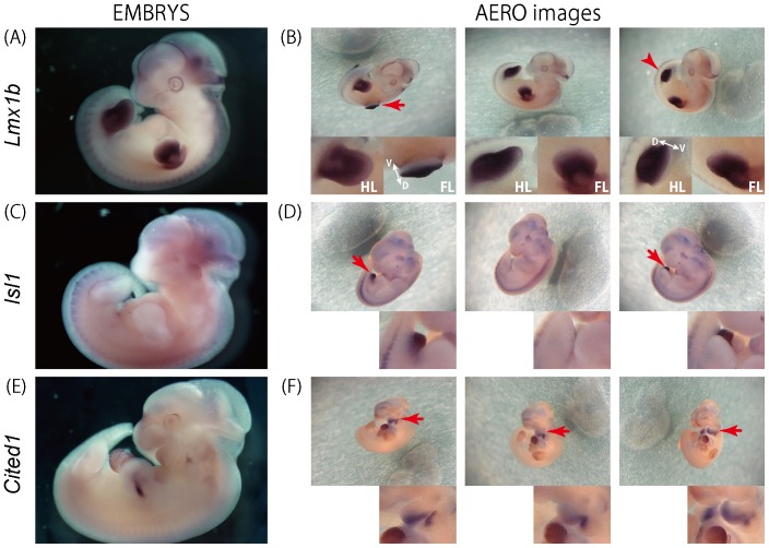
We have recently constructed a web-based database of gene expression in the mouse whole embryo, EMBRYS (http://embrys.jp/embrys/html/MainMenu.html). To allow examination of gene expression patterns to the fullest extent possible, this database provides both photo images and annotation data. However, since embryos develop via an intricate process of morphogenesis, it would be of great value to track embryonic gene expression from a three dimensional perspective. In fact, several methods have been developed to achieve this goal, but highly laborious procedures and specific operational skills are generally required. We utilized a novel microscopic technique that enables the easy capture of rotational, 3D-like images of the whole embryo. In this method, a rotary head equipped with two mirrors that are designed to obtain an image tilted at 45 degrees to the microscope stage captures serial images at 2-degree intervals. By a simple operation, 180 images are automatically collected. These 2D images obtained at multiple angles are then used to reconstruct 3D-like images, termed AERO images. By means of this system, over 800 AERO images of 191 gene expression patterns were captured. These images can be easily rotated on the computer screen using the EMBRYS database so that researchers can view an entire embryo by a virtual viewing on a computer screen in an unbiased or non-predetermined manner. The advantages afforded by this approach make it especially useful for generating data viewed in public databases.
Conflict of interest statement
Figures
References
-
- Kim TH, Ren B (2006) Genome-wide analysis of protein-DNA interactions. Annu Rev Genomics Hum Genet 7: 81–102. - PubMed
-
- Visel A, Thaller C, Eichele G (2004) GenePaint.org: an atlas of gene expression patterns in the mouse embryo. Nucleic Acids Res 32: D552–556. - PMC - PubMed
-
- Lein ES, Hawrylycz MJ, Ao N, Ayres M, Bensinger A, et al. (2007) Genome-wide atlas of gene expression in the adult mouse brain. Nature 445: 168–176. - PubMed
Publication types
MeSH terms
LinkOut - more resources
Full Text Sources
Other Literature Sources
Molecular Biology Databases
