. 2013 Oct 11;8(10):e75976.
doi: 10.1371/journal.pone.0075976. eCollection 2013.

A novel branched chain amino acids responsive transcriptional regulator, BCARR, negatively acts on the proteolytic system in Lactobacillus helveticus

Affiliations

A novel branched chain amino acids responsive transcriptional regulator, BCARR, negatively acts on the proteolytic system in Lactobacillus helveticus

Taketo Wakai et al. PLoS One. .

Abstract

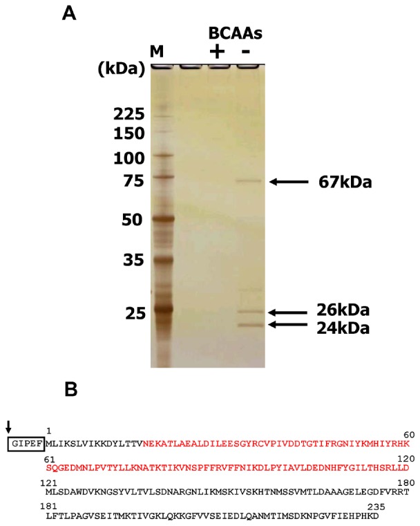
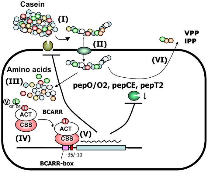
Transcriptional negative regulation of the proteolytic system of Lactobacillus helveticus CM4 in response to amino acids seems to be very important for the control of antihypertensive peptide production; however, it remains poorly understood. A 26-kDa protein with N-terminal cystathionine β-synthase domains (CBS domain protein), which seems to be involved in the regulatory system, was purified by using a DNA-sepharose bound 300-bp DNA fragment corresponding to the upstream regions of the six proteolytic genes that are down-regulated by amino acids. The CBS domain protein bound to a DNA fragment corresponding to the region upstream of the pepV gene in response to branched chain amino acids (BCAAs). The expression of the pepV gene in Escherichia coli grown in BCAA-enriched medium was repressed when the CBS domain protein was co-expressed. These results reveal that the CBS domain protein acts as a novel type of BCAA-responsive transcriptional regulator (BCARR) in L. helveticus. From comparative analysis of the promoter regions of the six proteolysis genes, a palindromic AT-rich motif, 5'-AAAAANNCTWTTATT-3', was predicted as the consensus DNA motif for the BCARR protein binding. Footprint analysis using the pepV promotor region and gel shift analyses with the corresponding short DNA fragments strongly suggested that the BCARR protein binds adjacent to the pepV promoter region and affects the transcription level of the pepV gene in the presence of BCAAs. Homology search analysis of the C-terminal region of the BCARR protein suggested the existence of a unique βαββαβ fold structure that has been reported in a variety of ACT (aspartate kinase-chorismate mutase-tyrA) domain proteins for sensing amino acids. These results also suggest that the sensing of BCAAs by the ACT domain might promote the binding of the BCARR to DNA sequences upstream of proteolysis genes, which affects the gene expression of the proteolytic system in L. helveticus.


Conflict of interest statement

Competing Interests: The two authors were employed by a commercial company (CALPIS Co., Ltd.). This does not alter the authors’ adherence to all the PLOS ONE policies on sharing data and materials.

Figures

Figure 1
Figure 1. Affinity purified proteins from L. helveticus CM4 eluted from DNA-sepharose.
DNA binding proteins purified by DNA-sepharose, which was bound to an approximately 300 bp DNA fragment corresponding to the upstream region of 6 proteolytic genes, were analyzed by SDS (5–15%)-PAGE as described in Materials and Methods (A). Proteins were eluted with buffer containing 10 mM BCAAs (Val, Leu, Ile) (+) or no BCAAs (−). The gel was silver stained after electrophoresis. The molecular masses of the marker proteins (lane M) are given on the left. Schematic drawing of the glutathione S-transferase (GST) fused 26 kDa cystathionine ß-synthase (CBS) domain protein is shown (B). The C-terminal GST protein is shown in the box. The arrow indicates the cleavage site for Factor Xa. Red letters indicate two tandem CBS domains (CBS pair) in the 26 kDa protein.
Figure 2
Figure 2. Electrophoresis mobility shift assay showing DNA binding.
Electrophoresis mobility shift assay using the purified CBS domain protein (CBSDP) and a 309 bp DNA fragment from upstream of the pepV gene in the presence or absence of 10 mM BCAAs and various amounts of CBS domain protein.
Figure 3
Figure 3. Transcriptional regulation by the CBS domain protein.
Schematic drawing of the expression plasmid (A). The pepV gene including 500 bp of upstream sequence was expressed in E. coli HB101 cells carrying pBR-pepV. The gene encoding the CBS domain protein was also co-expressed with the pepV gene in E. coli HB101 harboring pBR-pepV-CBS. White boxes show about 500 bp of DNA upstream of the genes. CBSDP and pepV indicate the ORF of each gene. pepV gene transcription in the E. coli transformant was quantified by RT-PCR with total RNA from E. coli cultured with or without 0.4% casamino acids and 10 mM BCAAs (B). The glyceraldehyde 3-phosphate dehydrogenase (GAPDH) gene (gapRTF, gapRTR) was used as the internal reference. Error bars indicate standard deviations. Statistical analysis of the data from triplicate experiments was conducted using the Student’s t test. ***P<0.001.
Figure 4
Figure 4. The search for a consensus motif for BCARR binding.
Electrophoresis mobility shift assays with the promoter regions of proteolysis genes using the purified BCARR protein (A). The upstream regions of the pepT2, pepCE, pepO, pepO2 and dppD genes were incubated alone or with 2.2 µM BCARR protein and 10 mM BCAAs. A conserved motif was identified for these six upstream sequences by MEME analysis (B). The weight matrix shows the frequency of A, C, T, or G nucleotides (as indicated in the legend) at each position of the motif. The inversely repeated consensus sequence (indicated by arrows) deduced from these frequencies is shown below the diagram. W can be either A or T. N can be any nucleotide. A graphical representation of the identified motif was obtained at the Weblogo website (http://weblogo.berkeley.edu/logo.cgi). Nucleotide positions are relative to the start codon of each proteolysis gene or the first gene in each operon (pepCE and pepO).
Figure 5
Figure 5. Electrophoresis mobility shift assay with various DNA fragments of the region upstream of the pepV.
Mobility shift assay with or without 2.3 µM BCARR protein and 10 mM BCAAs (A). Each 2,500 cpm of 32P labeled DNA fragment (0.3 ng) was incubated as described in Materials and Methods. Protein-DNA complexes were analyzed by 10% polyacrylamide gel electrophoresis. The 32P signals in the gel were detected by exposure to X-ray film. The position of the shifted bands is indicated in the right margin by arrows (weak shift indicated by a small arrow, and a significant shift by a big arrow; the origin is indicated by the open arrow). DNA fragments used in the above experiment and the results of the EMSA are summarized in (B). Nucleotide positions are relative to the start codon. BCARR-Box indicates the predicted BCARR binding motif by MEME analysis. ORF shows the open reading frame of the pepV gene. The intensity of the shifted bands is indicated by + (slight probe shifted) to +++ (all probes shifted).
Figure 6
Figure 6. DNase I footprinting analysis and schematic drawing of the BCARR protein-DNA binding.
Single-end (forward 5′strand) radioactively labeled probes containing 290 bp of sequence upstream of pepV were examined alone (lane 1) or with 2.3 µM (lane 2) and with 18 µM (lane 3) BCARR protein (A). Lane AG; A+G ladder prepared from the labelled DNA as described in Materials and Methods. The numbers at the left indicate the nucleotide number from the start codon. The vertical bar at the right represents the protected region. Solid lines and dotted lines indicate strongly and weakly protected regions, respectively. Schematic drawing of the DNaseI footprint analysis (B). The binding site of the BCARR protein was located in the sequence upstream of the pepV gene. The consensus motif detected in the upstream sequences of the six proteolysis genes ranging from −91 to −77 in pepV gene is shown by a yellow box. The putative promoters, −35 (TTGAAA) and −10 (TATTTT), are underlined and in blue letters. The nucleotide numbers indicate the distance from the start codon.
Figure 7
Figure 7. Homology search and sequence alignment of the ACT domain of the L. helveticus BCARR protein.
Predicted schematic domain structure of the L. helveticus BCARR protein and proteins with similarity to the C-terminal region of the BCARR identified by PSI-BLAST (A). Sequence alignment of the ACT domain of the L. helveticus BCARR protein and reported ACT domain proteins (B). The sequence alignment was performed using ClustalW. Helices and beta-strands are represented by arrows and are labeled “α” and “β” respectively. The color scheme generally follows that of Grant et al. , and Aravind and Koonin and refers to the following residue types: green, hydrophobic (ILVCAGMFYWTP); magenta, polar (HKREQDNST); gray, large (FILMWYKREQ); yellow, small (ACGSTDNVP); and red, conserved glycine. IlvH; Acetohydroxylate synthase small regulatory subunit [Nitrosomonas europaea] (ref|NC_004757.1|), ASSS; Acetolactate synthase small subunit [Synechococcus sp. WH 7803] (emb|CAK24070.1|), TDH; threonine dehydratase [Selenomonas noxia F0398] (gb|EHG23294.1|), AUP; acetoin utilization protein [Lysinibacillus sphaericus C3-41] (ref|YP_001699789.1|). Symbols “ * ”, “ : ” and “. ” are shown according to the method of ClustralW. ‘*’ indicates positions are completely conserved. ‘:’ indicates a fully conserved ‘strong’ group. ‘.’ indicates one of the fully conserved ‘weaker’ groups.
Figure 8
Figure 8. The phylogenetic tree of the BCARR.
Homologous protein sequences were identified by BLASTP searches (cutoff e-value >10−50). An unrooted phylogenetic tree displaying branch lengths that was built using ClustalW and the NJ algorithm is shown. To simplify the tree, one sequence was selected from each genus. For the genus Lactobacillus, four sequences were selected from the delbruekii subgroup including L. helveticus, one sequence from each subgroup other than the delbruekii subgroup as defined in the previous report . The scale bar represents base changes per site. A. : Alloiococcus, B. : Bacillus, D. : Dolosigranulum, E. : Enterococcus, F. : Fructobacillus, G. : Granulicatella, Ls. : Leuconostoc, Li. : Listeria, M. : Melissococcus, P. : Pediococcus, T. : Tetragenococcus, W. : Weissella. * Carnobacteriaceae also includes G. adiacens.
Figure 9
Figure 9. Model for the regulation of the proteolytic system of L. helveticus by the BCARR protein.
Release of the long peptide containing VPP and IPP sequences by an extracellular proteinase (I), uptake of the long peptide via oligopeptide transporter (II), intracellular processing of the peptides to amino acids by peptidases (III), BCARR (ACT domain)-BCAA complex formation (IV), binding of the complex (CBS domain) to the BCARR-box and repression of proteolysis gene transcription (V) and repressed release of the antihypertensive peptides, VPP and IPP (VI). L; Leucine, I; Isoleucine, V; Valine. T arrows indicate repressive effects.

Similar articles

Cited by

References

    1. Slattery L, O’Callaghan J, Fitzgerald G, Beresford T, Ross R (2010) Invited review: Lactobacillus helveticus–A thermophilic dairy starter related to gut bacteria. Journal of dairy science 93: 4435–4454. - PubMed
    1. Gatti M, Trivisano C, Fabrizi E, Neviani E, Gardini F (2004) Biodiversity among Lactobacillus helveticus strains isolated from different natural whey starter cultures as revealed by classification trees. Applied and environmental microbiology 70: 182–190. - PMC - PubMed
    1. Sneath PH, Mair NS, Sharpe ME, Holt JG (1986) Bergey’s manual of systematic bacteriology. Volume 2: Williams & Wilkins.
    1. Sadat-Mekmene L, Genay M, Atlan D, Lortal S, Gagnaire V (2011) Original features of cell-envelope proteinases of Lactobacillus helveticus. A review. International Journal of Food Microbiology 146: 1–13. - PubMed
    1. Yamamoto N, Akino A, Takano T (1994) Antihypertensive effects of different kinds of fermented milk in spontaneously hypertensive rats. Bioscience, biotechnology, and biochemistry 58: 776–778.

MeSH terms

LinkOut - more resources