

Expression of Tau40 induces activation of cultured rat microglial cells
- PMID: 24146816
- PMCID: PMC3795725
- DOI: 10.1371/journal.pone.0076057
Expression of Tau40 induces activation of cultured rat microglial cells
Abstract
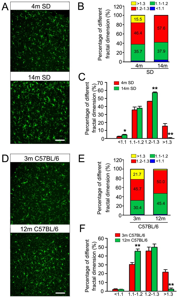
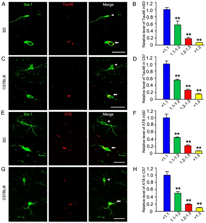
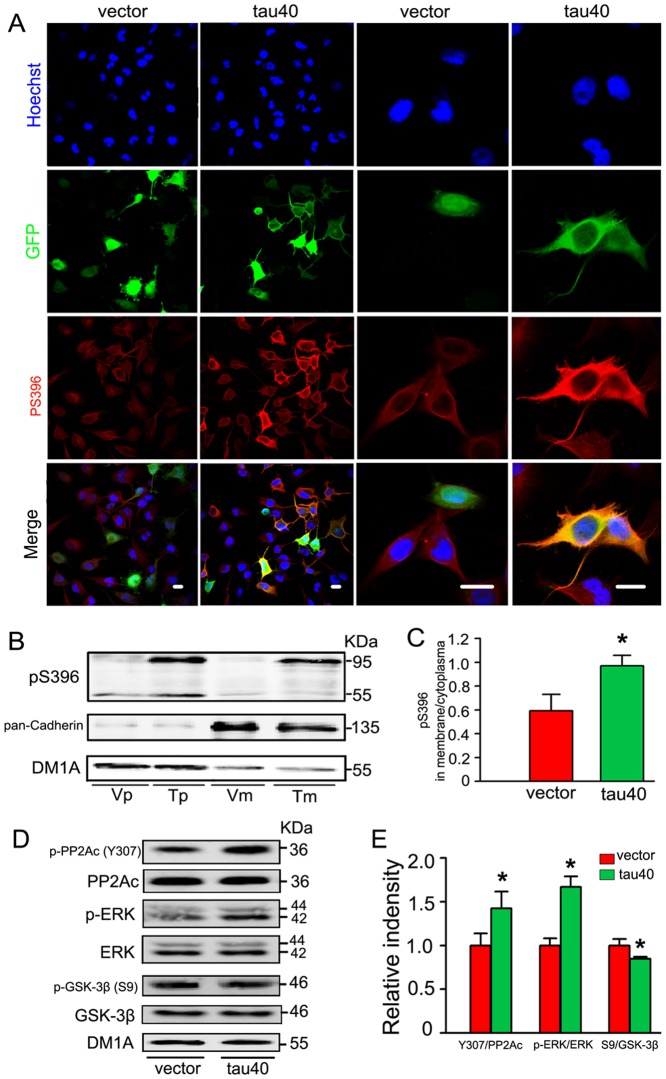
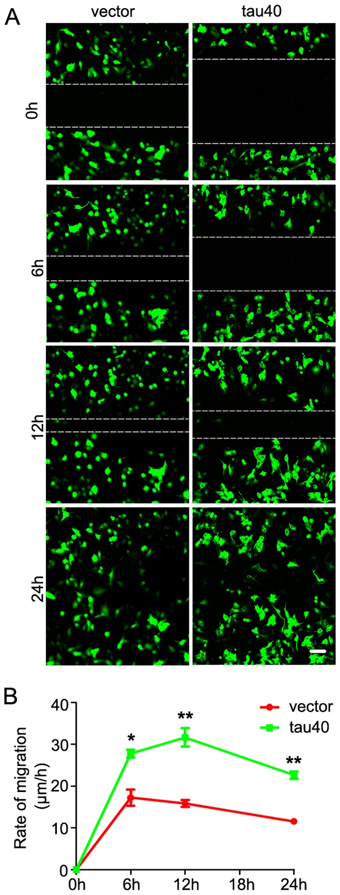
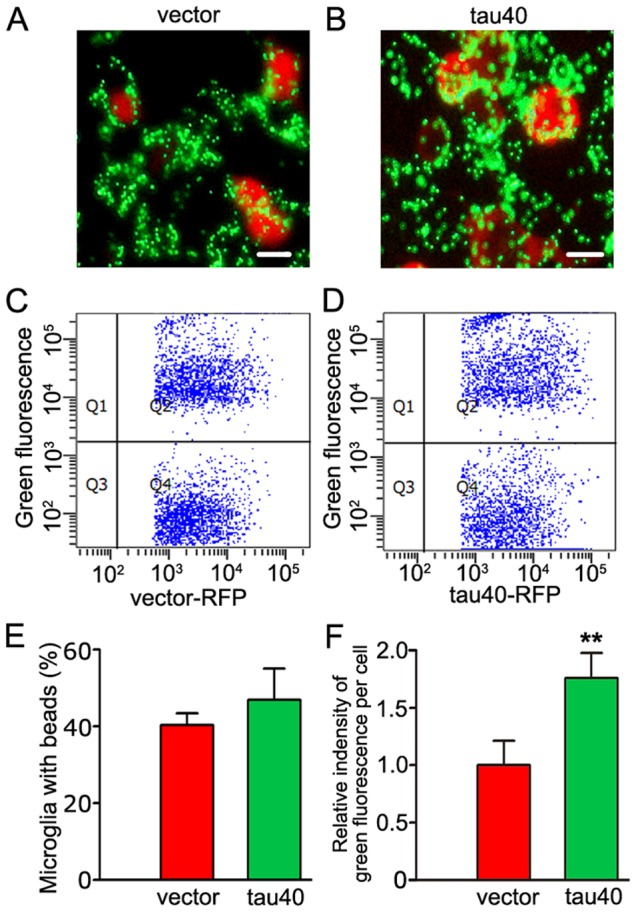
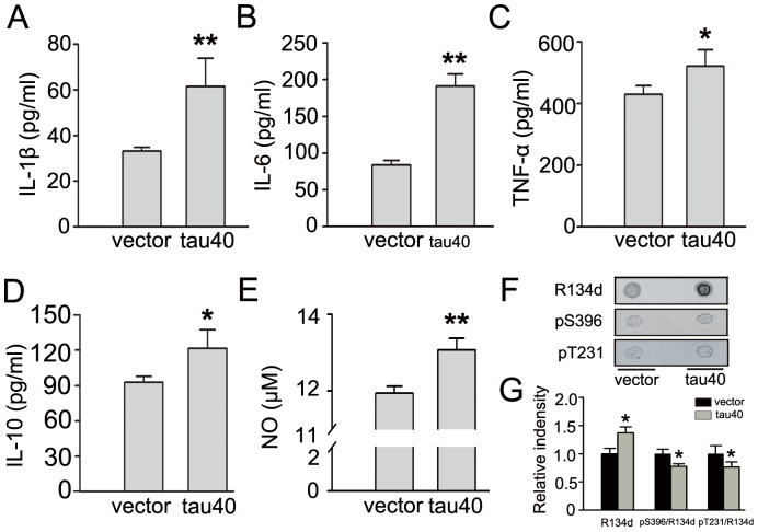
Accumulation of microtubule-associated protein tau has been observed in the brain of aging and tauopathies. Tau was observed in microglia, but its role is not illustrated. By immunofluorescence staining and the fractal dimension value assay in the present study, we observed that microglia were activated in the brains of rats and mice during aging, simultaneously, the immunoreactivities of total tau and the phosphorylated tau were significantly enhanced in the activated microglia. Furtherly by transient transfection of tau40 (human 2N/4R tau) into the cultured rat microglia, we demonstrated that expression of tau40 increased the level of Iba1, indicating activation of microglia. Moreover, expression of tau40 significantly enhanced the membranous localization of the phosphorylated tau at Ser396 in microglia possibly by a mechanism involving protein phosphatase 2A, extracellular signal-regulated kinase and glycogen synthase kinase-3β. It was also found that expression of tau40 promoted microglial migration and phagocytosis, but not proliferation. And we observed increased secretion of several cytokines, including interleukin (IL)-1β, IL-6, IL-10, tumor necrosis factor-α and nitric oxide after the expression of tau40. These data suggest a novel role of human 2N/4R tau in microglial activation.
Conflict of interest statement
Figures

Similar articles
-
FBXW5 reduction alleviates spinal cord injury (SCI) by blocking microglia activity: A mechanism involving p38 and JNK.Biochem Biophys Res Commun. 2019 Jun 25;514(2):558-564. doi: 10.1016/j.bbrc.2019.04.086. Epub 2019 May 3. Biochem Biophys Res Commun. 2019. PMID: 31060780
-
Micheliolide suppresses LPS-induced neuroinflammatory responses.PLoS One. 2017 Oct 17;12(10):e0186592. doi: 10.1371/journal.pone.0186592. eCollection 2017. PLoS One. 2017. PMID: 29040306 Free PMC article.
-
Toll-like receptor-9 (TLR-9) deficiency alleviates optic nerve injury (ONI) by inhibiting inflammatory response in vivo and in vitro.Exp Cell Res. 2020 Nov 1;396(1):112159. doi: 10.1016/j.yexcr.2020.112159. Epub 2020 Jul 9. Exp Cell Res. 2020. PMID: 32652081
-
Beyond Activation: Characterizing Microglial Functional Phenotypes.Cells. 2021 Aug 28;10(9):2236. doi: 10.3390/cells10092236. Cells. 2021. PMID: 34571885 Free PMC article. Review.
-
G-protein coupled purinergic P2Y12 receptor interacts and internalizes TauRD-mediated by membrane-associated actin cytoskeleton remodeling in microglia.Eur J Cell Biol. 2022 Apr;101(2):151201. doi: 10.1016/j.ejcb.2022.151201. Epub 2022 Jan 25. Eur J Cell Biol. 2022. PMID: 35101770 Review.
Cited by
-
Phagocytosis of full-length Tau oligomers by Actin-remodeling of activated microglia.J Neuroinflammation. 2020 Jan 8;17(1):10. doi: 10.1186/s12974-019-1694-y. J Neuroinflammation. 2020. PMID: 31915009 Free PMC article.
-
Microglia in neurodegenerative diseases: mechanism and potential therapeutic targets.Signal Transduct Target Ther. 2023 Sep 22;8(1):359. doi: 10.1038/s41392-023-01588-0. Signal Transduct Target Ther. 2023. PMID: 37735487 Free PMC article. Review.
-
Associations between brain inflammatory profiles and human neuropathology are altered based on apolipoprotein E ε4 genotype.Sci Rep. 2020 Feb 19;10(1):2924. doi: 10.1038/s41598-020-59869-5. Sci Rep. 2020. PMID: 32076055 Free PMC article.
-
Expression of 1N3R-Tau isoform inhibits cell proliferation by inducing S phase arrest in N2a cells.PLoS One. 2015 Mar 30;10(3):e0119865. doi: 10.1371/journal.pone.0119865. eCollection 2015. PLoS One. 2015. PMID: 25822823 Free PMC article.
-
Molecular and cellular mechanisms underlying the pathogenesis of Alzheimer's disease.Mol Neurodegener. 2020 Jul 16;15(1):40. doi: 10.1186/s13024-020-00391-7. Mol Neurodegener. 2020. PMID: 32677986 Free PMC article. Review.
References
-
- Vaughan DW, Peters A (1974) Neuroglial cells in the cerebral cortex of rats from young adulthood to old age: an electron microscope study. J Neurocytol 3: 405–429. - PubMed
-
- Becher B, Prat A, Antel JP (2000) Brain-immune connection: immuno-regulatory properties of CNS-resident cells. Glia 29: 293–304. - PubMed
-
- Aloisi F (2001) Immune function of microglia. Glia 36: 165–179. - PubMed
-
- McGeer PL, Rogers J, McGeer EG (2006) Inflammation, anti-inflammatory agents and Alzheimer disease: the last 12 years. J Alzheimers Dis 9: 271–276. - PubMed
Publication types
MeSH terms
Substances
LinkOut - more resources
Full Text Sources
Other Literature Sources
Miscellaneous
