. 2013 Oct 16;8(10):e76115.
doi: 10.1371/journal.pone.0076115. eCollection 2013.

Expression of the B-cell receptor component CD79a on immature myeloid cells contributes to their tumor promoting effects

Affiliations

Expression of the B-cell receptor component CD79a on immature myeloid cells contributes to their tumor promoting effects

Dror Luger et al. PLoS One. .

Abstract

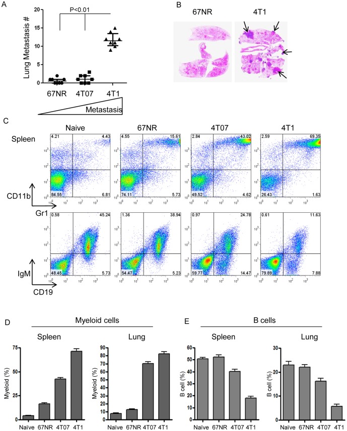
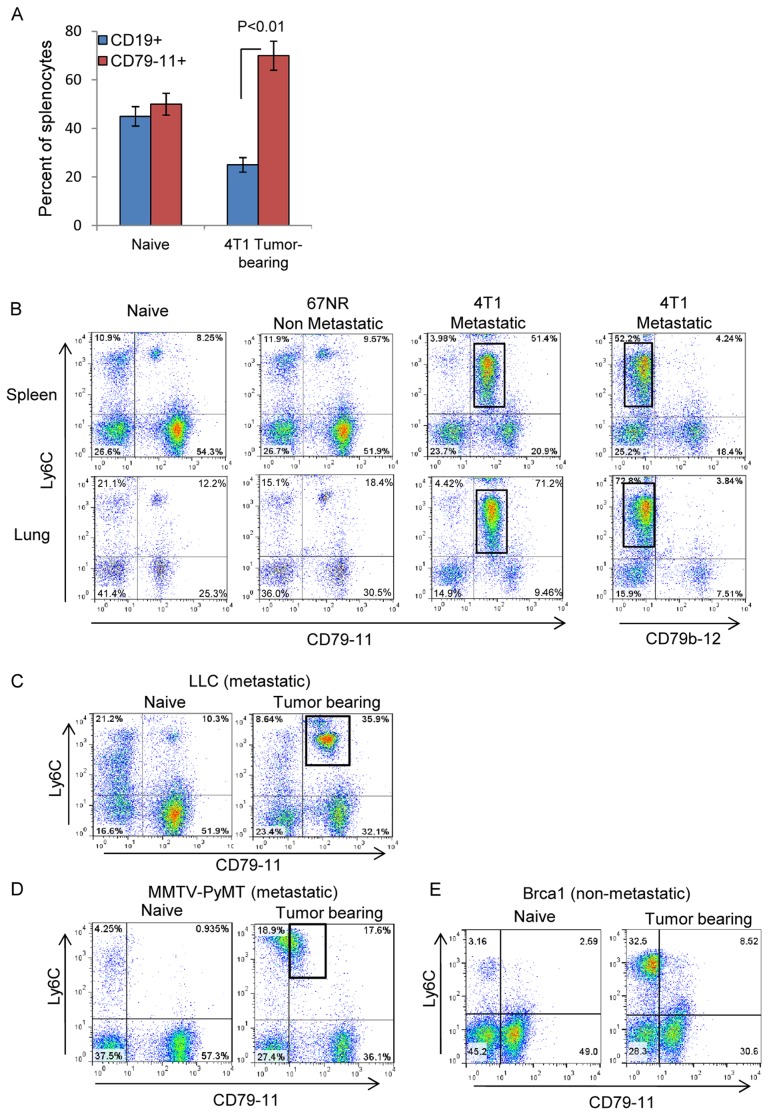
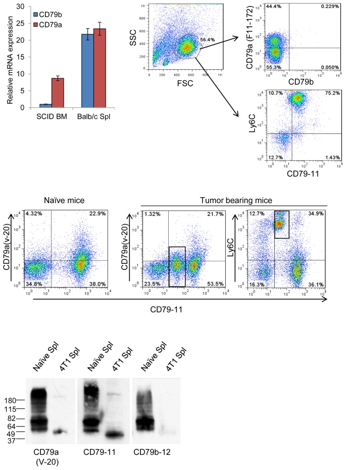
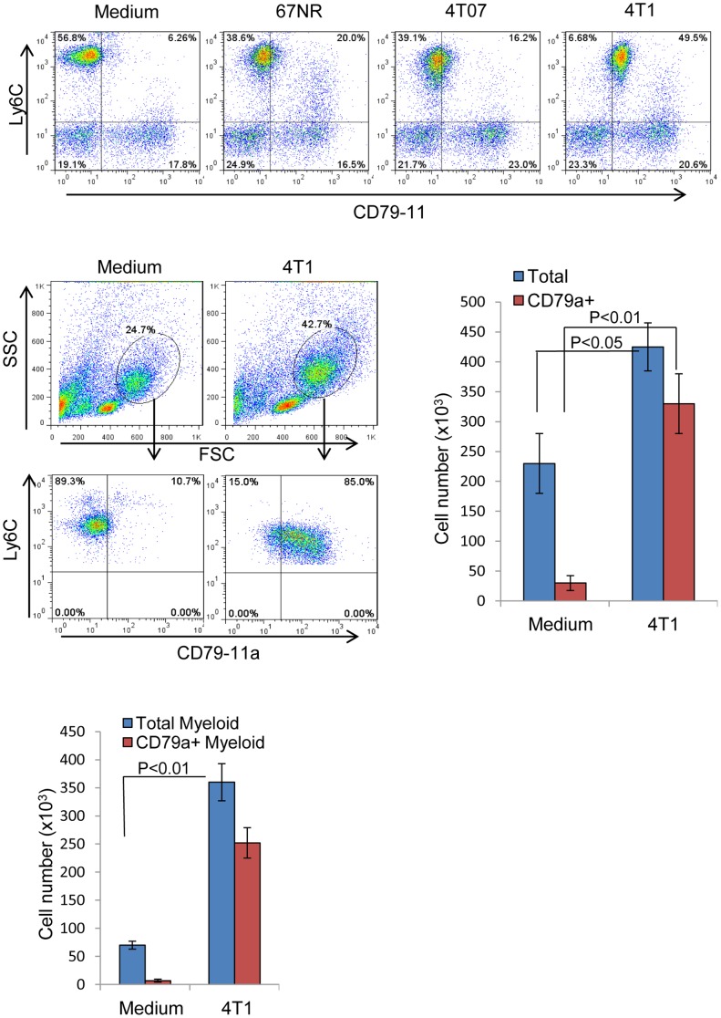
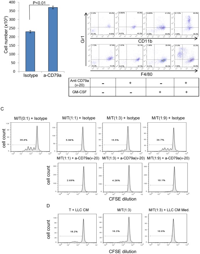
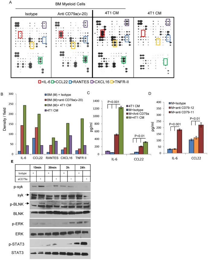
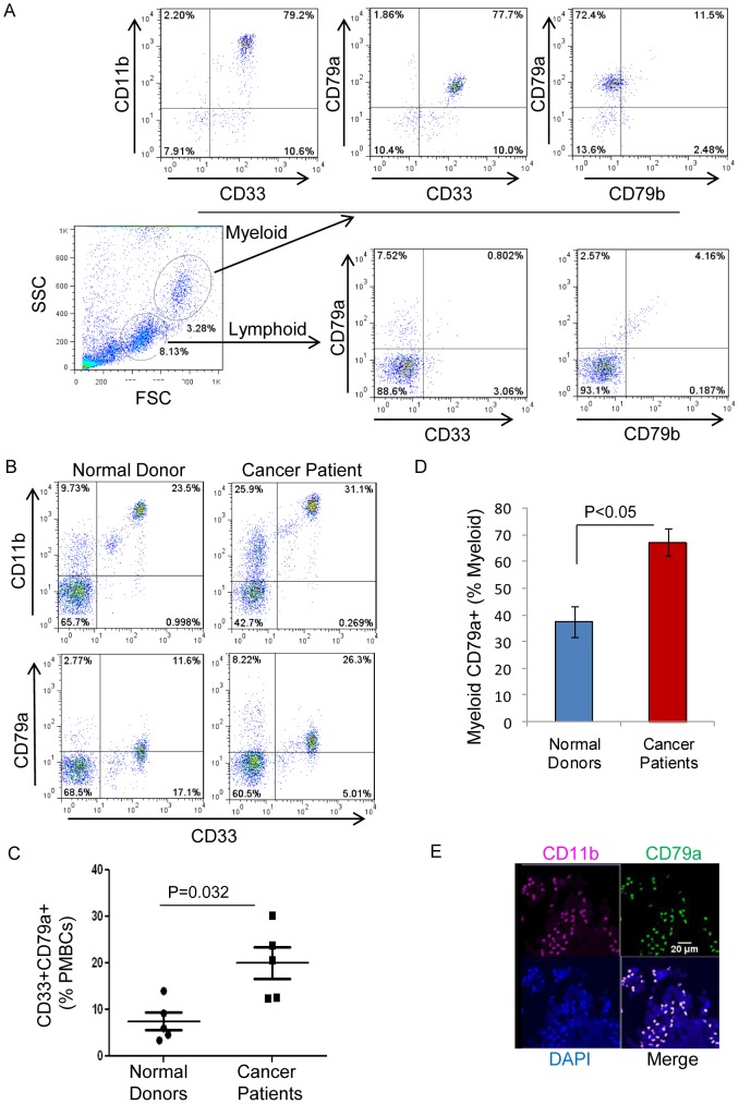
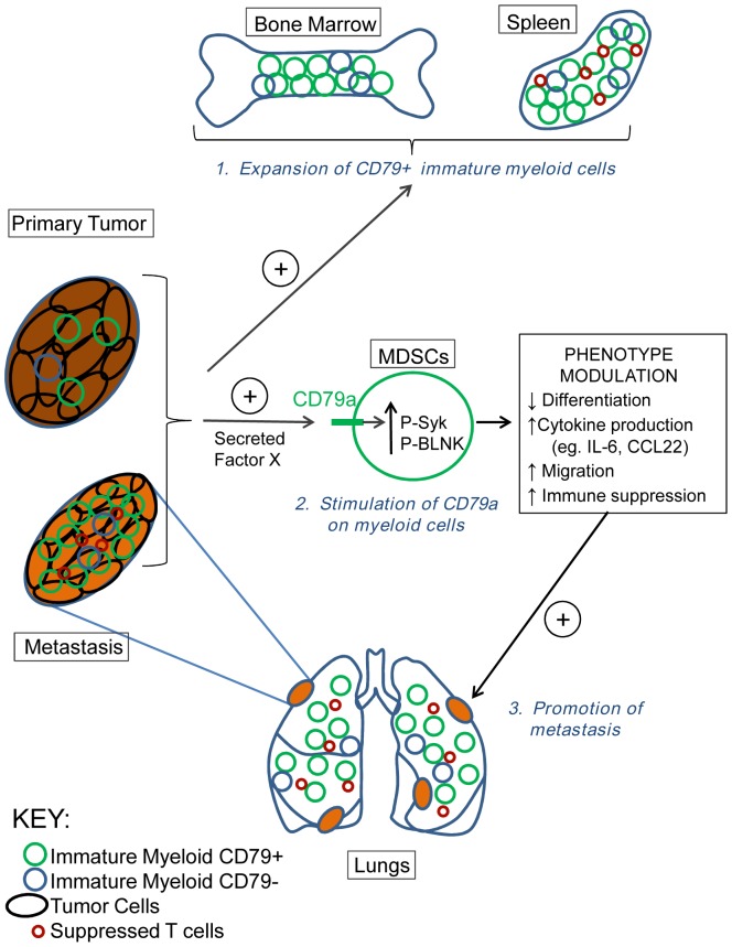
The role of myeloid derived suppressor cells (MDSCs) in promoting tumorigenesis is well-established, and significant effort is being made to further characterize surface markers on MDSCs both for better diagnosis and as potential targets for therapy. Here we show that the B cell receptor adaptor molecule CD79a is unexpectedly expressed on immature bone marrow myeloid cells, and is upregulated on MDSCs generated in multiple different mouse models of metastatic but not non-metastatic cancer. CD79a on MDSCs is upregulated and activated in response to soluble factors secreted by tumor cells. Activation of CD79a on mouse MDSCs, by crosslinking with a specific antibody, maintained their immature phenotype (CD11b+Gr1+), enhanced their migration, increased their suppressive effect on T cell proliferation, and increased secretion of pro-tumorigenic cytokines such as IL-6 and CCL22. Furthermore, crosslinking CD79a on myeloid cells activated signaling through Syk, BLNK, ERK and STAT3 phosphorylation. In vivo, CD79+ myeloid cells showed enhanced ability to promote primary tumor growth and metastasis. Finally we demonstrate that CD79a is upregulated on circulating myeloid cells from lung cancer patients, and that CD79a+ myeloid cells infiltrate human breast tumors. We propose that CD79a plays a functional role in the tumor promoting effects of myeloid cells, and may represent a novel target for cancer therapy.


Conflict of interest statement

Competing Interests: The authors have declared that no competing interests exist.

Figures

Figure 1
Figure 1. Increased myeloid cells and reduced B-cells associated with increasing metastatic efficiency in breast cancer models.
Metastatic 4T1 and poorly- or non-metastatic 4T07 and 67NR cell lines were implanted orthotopically in Balb/c mice, and the extent of lung metastasis and the relative representation of immature myeloid (CD11b+Gr1+) and mature B (CD19+IgM+) cells in lungs and spleen were analyzed 28 days post-inoculation. (A) Number of histologically-detectable metastases in lungs from mice inoculated with the different cell lines. N = 8 mice/group. Bars show median and interquartile range. (B) Representative histological cross-sections of lungs from mice with 67NR or 4T1 tumors. Arrows indicate metastases. (C) Representation of immature myeloid cells (CD11b+Gr1+) and mature B cells (IgM+CD19+) in spleens from the different groups as assessed by flow cytometry. (D) Histograms summarizing relative numbers of immature myeloid cells and mature B-cells as a percent of viable leukocytes in naïve and tumor-bearing mice. N = 5 mice/group.
Figure 2
Figure 2. Expression of the B-cell receptor subunit CD79a on MDSCs induced by metastatic tumors.
CD79a/b expression on the Ly6C+ myeloid cell population in multiple tumor models was assessed by flow cytometry. (A) Expression of the B-cell markers CD19 and CD79a/b on splenocytes from naïve and 67NR and 4T1 tumor-bearing mice. (B) Ly6C and CD79a/b expression on viable leukocytes from lungs and spleens of Balb/c mice bearing 67NR or 4T1 tumors at day 28 post-inoculation. The box indicates the immature myeloid population. CD79 on myeloid cells is recognized by the CD79-11 but not the CD79-12 antibody. (C) CD79a expression on MDSCs was also evaluated in the Lewis Lung Carcinoma transplantable metastatic lung cancer model (C57Bl/6 background). (D,E) Similar evaluation was done in two genetically-engineered models of breast cancer; the metastatic MMTV-PyMT model (D) on the FVB/N background (age 3 months) and the non-metastatic Brca1 model (E) on a mixed genetic background (age 6 months).
Figure 3
Figure 3. Expression of CD79a mRNA and protein in bone marrow cells from B-cell deficient mice.
(A) RNA was extracted from BM cells of naïve SCID mice and from spleen of naïve Balb/c mice and CD79a and CD79b mRNA expression were measured by qRT-PCR. RNA levels were normalized to PPIAand presented relative to the level of CD79b mRNA in SCID BM. Results are mean +/− SEM; n = 3. (B) BM-derived leukocytes from naïve SCID mice were analyzed by flow cytometry for intracellular epitopes of CD79a using the anti-mouse CD79a clone F11-172. (C) To assess the extracellular expression of CD79a, BM cells from mice bearing LLC tumors were co-stained with anti CD79-11 together with the polyclonal anti-CD79a(v-20), which showed a weaker pattern of staining than CD79-11. Boxes indicate the myeloid cells. (D) CD79a/b protein in splenocytes from naïve and 4T1 tumor-bearing mice was assessed by western blot under reducing conditions. Data shown are representative of three independent experiments.
Figure 4
Figure 4. Tumor cell-secreted factors upregulate CD79a on myeloid cells and induce their expansion and migration.
(A) The ability of tumor cells of varying metastatic potential to induce expression of CD79a on immature myeloid cells from bone marrow of naïve mice was assessed using a Transwell® system (0.4 µm pore size) that prevented contact between tumor and bone marrow-derived cells. BM cells were co-incubated with tumor cells of medium alone for 48 h and then BM cells were analyzed by flow cytometry. (B) Representative FACS plots and histograms showing the effect of 4T1 secreted factors on the expansion of BM myeloid cells expressing CD79a, tested using a Transwell® system as in A. (C) Migration of BM cells in response to 4T1 secreted factors was tested in a Transwell® system as above, but using a 3.0 µm pore-size membrane to allow migration of BM cells towards the 4T1 cells in the well below. Cells that migrated through the membrane into the well below were collected, counted and analyzed by FACS for CD79a expression. Results are mean +/- SEM; n = 4 determinations.
Figure 5
Figure 5. CD79a activation on myeloid cells affects migration, differentiation and T-cell suppression.
(A) The effect of stimulation with anti CD79a(v-20) on the migration of FACS-sorted Ly6C+ immature myeloid cells from naive bone marrow was tested in a Transwell® system for 48 h. Results are mean +/− SEM; n = 3 determinations. (B) Bone marrow cells from naïve SCID mice were stimulated with anti- CD79a(v-20) (5 µg/ml) for 96 h with or without added GM-CSF in low (2%) normal mouse serum. The effect of anti CD79a(v-20) on myeloid cell differentiation status was assessed using immature myeloid (CD11b+Gr1+), granulocytic (Gr1+) and macrophage (F4/80+) markers. Data shown are representative of two independent experiments. (C) The effect of immature myeloid cells stimulated with anti CD79a on proliferation of CD4 T cells. Immunopurified naïve CD4+ T cells (“T”) labeled with CSFE were stimulated with anti CD3/CD28 with the addition of the indicated ratios of FACS-sorted immature BM myeloid cells (“M”) with or without the addition of anti CD79a(v-20) antibody. Cell divisions were measured by flow cytometry analysis for CFSE dilution. Dose response for effect on T-cell division of myeloid cells added at the indicated ratio, with the addition of anti CD79a(v-20) or isotype-matched control antibody is shown. (D) The effect of LLC tumor cell conditioned medium (CM) (ratio 1/5 v/v), myeloid cells and the combination of both on T cells proliferation. Data shown are representative of two independent experiments.
Figure 6
Figure 6. CD79a activation on myeloid cells alters cytokine expression and activates signaling pathways.
The secretion of cytokines and chemokines by Ly6C+ immature BM myeloid cells isolated from SCID mice and cultured for 48 h under the indicated conditions was assessed using a membrane-based cytokine array. (A) Arrays of conditioned media from culture of BM cells treated with anti- CD79a(v-20), isotype control, or 4T1 tumor cell CM, together with a control array for 4T1 CM alone. (B) Relative levels of proteins with the most significant change (indicated by boxes in A) were quantitated by densitometry. (C, D) Validation of changes in IL-6 and CCL22 by Quantikine ELISA. Both the arrays and the ELISA are representative of 2 independent experiments. (E) Induction of downstream signaling events triggered by cross-linking CD79a with anti CD79a(v-20) on BM cells from SCID mice was assessed by western blot.
Figure 7
Figure 7. CD79a expression and activation on myeloid cells contributes to their pro-tumorigenic effects in vivo.
(A) Immunofluorescence of normal lung and metastasis-bearing lungs in the LLC model. Infiltrating Gr1+CD79a+ myeloid cells are clearly visible at the outer edge of a lung metastasis but are rare or absent from normal lung tissue. The dotted line marks the outer boundary of the metastasis. Scale bar: 40 µm. (B) Quantification of lung infiltrating Gr1+CD79a+ and Gr1+Cd79-11- myeloid cells: Analysis was done for 3 groups: normal lung, uninvolved lung tissue from metastasis bearing lung, and lung metastasis. Each data point represents the mean of 5 fields (500 µm2) for a given sample and results are given as mean +/− SEM. (C) Immature BM cells from C57Bl/6 mice were FACS sorted into two groups of Ly6C+CD79a+ and Ly6C+ CD79a myeloid cells. The sorted myeloid cells were co-inoculated together with LLC tumor cells subcutaneously in C57Bl/6 mice at a ratio of 25∶1 myeloid: tumor cells. Tumor weight was measured at day 14 post-inoculation. Results are mean +/− SEM (n = 5). (D) Ly6C+ BM-derived myeloid cells from naïve mice were isolated by FACS and cultured with anti CD79a(v-20) or isotype control antibodies for 24 h. Treated myeloid cells were then harvested and injected i.v. together with luciferase-expressing LLC cells (2×10⁶ myeloid cells and 104 LLC tumor cells/mouse) in C57Bl/6 mice. Metastatic burden was quantitated from in vivo luciferase signal at day 15 after implantation. Results are median with interquartile range (n = 10 mice/group).
Figure 8
Figure 8. CD79a expression on myeloid cells from normal human donors and in cancer patients.
(A) Expression of CD79a on immature myeloid cells (characterized as CD11b+CD33+) in BM from a normal human donor. (B) Representative FACS profiles of immature myeloid cells in peripheral blood (live leukocyte gate) from a normal donor and a lung cancer patient. (C) Immature myeloid cells (CD11b+CD33+) expressing CD79a in normal donors and lung cancer patients as a% of live leukocytes. Results are mean +/− SEM (n = 5). (D) Histogram showing proportion of CD79a+ myeloid cells relative to total myeloid cells. Results are mean +/− SEM (n = 5) (E) Representative immunofluorescence staining of infiltrating CD79a+ myeloid cells in a human invasive breast carcinoma. Scale bar; 20 µm.
Figure 9
Figure 9. Role for CD79a in reciprocal interactions between immature myeloid and tumor cells that promote tumorigenesis and metastasis.
Metastatic tumors induce the expansion of an immature myeloid cell population in bone marrow and spleen, and drive enrichment of a subpopulation that expresses elevated CD79a. An as yet unidentified CD79a ligand secreted by tumor cells activates the CD79a on the immature myeloid cells leading to signaling through Syk and BLNK and modulation of cellular phenotype in a variety of ways. These include maintainance of the immature state, upregulated expression of tumor-promoting cytokines and chemokines, increased migration, and enhanced immunosuppressive activity. This phenotypic modulation induces and/or amplifies the stimulatory effect of MDSCs on tumorigenesis and metastasis.

Similar articles

Cited by

References

    1. Gabrilovich DI, Nagaraj S (2009) Myeloid-derived suppressor cells as regulators of the immune system. Nat Rev Immunol 9: 162–174. - PMC - PubMed
    1. Sica A, Bronte V (2007) Altered macrophage differentiation and immune dysfunction in tumor development. J Clin Invest 117: 1155–1166. - PMC - PubMed
    1. Ostrand-Rosenberg S, Sinha P, Beury DW, Clements VK (2012) Cross-talk between myeloid-derived suppressor cells (MDSC), macrophages, and dendritic cells enhances tumor-induced immune suppression. Semin Cancer Biol 22: 275–281. - PMC - PubMed
    1. Yang L, DeBusk LM, Fukuda K, Fingleton B, Green-Jarvis B, et al. (2004) Expansion of myeloid immune suppressor Gr+CD11b+ cells in tumor-bearing host directly promotes tumor angiogenesis. Cancer Cell 6: 409–421. - PubMed
    1. Yan HH, Pickup M, Pang Y, Gorska AE, Li Z, et al. (2010) Gr-1+CD11b+ Myeloid Cells Tip the Balance of Immune Protection to Tumor Promotion in the Premetastatic Lung. Cancer Res 70: 6139–6149. - PMC - PubMed

Publication types

MeSH terms