. 2013 Oct 11;8(10):e76279.
doi: 10.1371/journal.pone.0076279. eCollection 2013.

A modular lentiviral and retroviral construction system to rapidly generate vectors for gene expression and gene knockdown in vitro and in vivo

Affiliations

A modular lentiviral and retroviral construction system to rapidly generate vectors for gene expression and gene knockdown in vitro and in vivo

Benjamin Geiling et al. PLoS One. .

Abstract

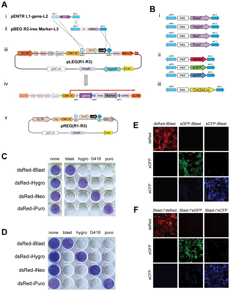
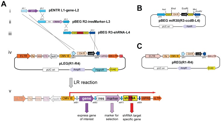
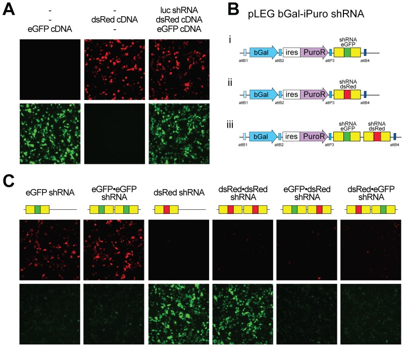
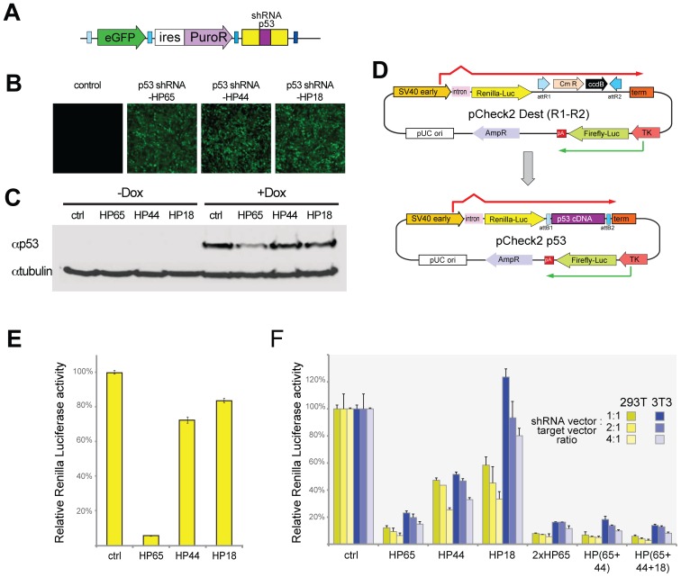
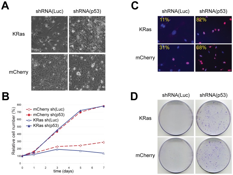
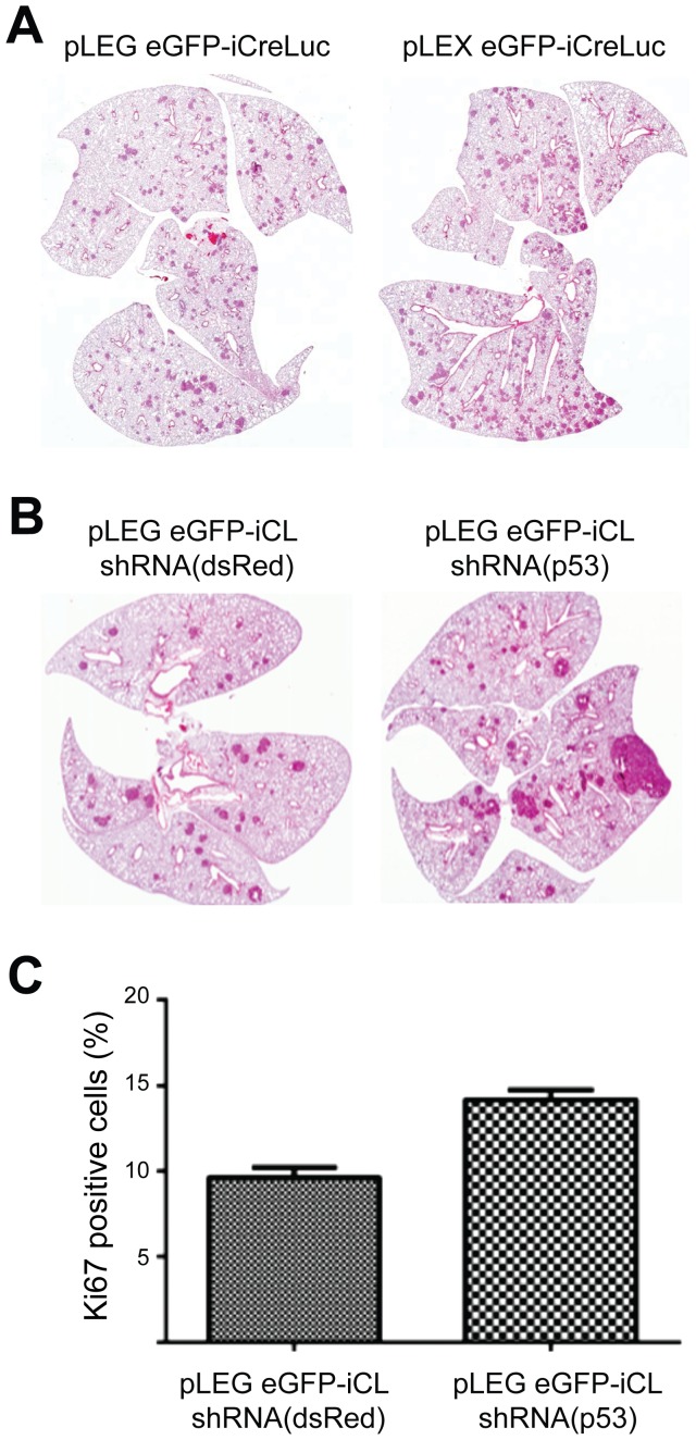
The ability to express exogenous cDNAs while suppressing endogenous genes via RNAi represents an extremely powerful research tool with the most efficient non-transient approach being accomplished through stable viral vector integration. Unfortunately, since traditional restriction enzyme based methods for constructing such vectors are sequence dependent, their construction is often difficult and not amenable to mass production. Here we describe a non-sequence dependent Gateway recombination cloning system for the rapid production of novel lentiviral (pLEG) and retroviral (pREG) vectors. Using this system to recombine 3 or 4 modular plasmid components it is possible to generate viral vectors expressing cDNAs with or without inhibitory RNAs (shRNAmirs). In addition, we demonstrate a method to rapidly produce and triage novel shRNAmirs for use with this system. Once strong candidate shRNAmirs have been identified they may be linked together in tandem to knockdown expression of multiple targets simultaneously or to improve the knockdown of a single target. Here we demonstrate that these recombinant vectors are able to express cDNA and effectively knockdown protein expression using both cell culture and animal model systems.


Conflict of interest statement

Competing Interests: The authors have declared that no competing interests exist.

Figures

Figure 1
Figure 1. Modular design and function of pLEG/pREG viral vector expression system.
A) A generalized three-plasmid LR recombination reaction depicting the insertion of a gene and selection marker into a lentiviral backbone. Each attLx site recombines with a corresponding attRx site and the order and orientation of these sites directs the formation of the recombinant attXx site as well as the insert order/orientation. AttL1-attL2 (i) and attR2-attL3 (ii) flanked entry vectors recombine with a lentiviral destination vector, pLEG(R1–R3) (iii) producing a recombinant lentiviral expression vector that when integrated contains a single CMV-driven bicistronic transcript (iv). Retroviral destination vectors (pREG) are also possible and function in the same manner (v). LTR: Long Terminal Repeat, Psi: packaging signal, RRE: Rev Response Element, CTS: central PolyPurine Tract, CMV IE: cytomegalovirus-immediate early, WPRE: Woodchuck hepatitis Post-transcriptional Regulatory Element, ΔLTR: Self Inactivated LTR. B) Drug resistance markers (i) for use with the pLEG/pREG system along with fluorophore markers (ii) and Cre2ALuc (iii) which may be inserted and expressed downstream of any attL1-attL2 flanked gene. C) Stable NIH 3T3 cell lines expressing each of the four drug resistant markers after infection by a recombinant lentiviral (pLEG) vector produced by three-plasmid recombination reaction. Giemsa staining highlights the drug resistant populations for each case. D) Stable NIH 3T3 cell lines expressing each of the four drug resistant markers after infection by a recombinant retroviral (pREG) vector – as in (C). E) Stable HEK 293T cell lines expressing each of the three upstream fluorophore markers after infection by a recombinant lentiviral (pLEG) vector produced by three-plasmid recombination reaction. F) Stable HEK 293T cell lines expressing each of the three downstream fluorophore markers – as in (E). Psi: RNA packaging symbol; SIN LTR: self-inactivating long terminal repeat; WPRE: Woodchuck hepatitis virus post-transcriptional element; CmR/ccdB: Chloramphenicol resistance/ccdB cell death cassette; ZeoR: Zeocin resistance cassette; pA: poly adenylation signal; AmpR: Ampicillin resistance gene; HygroR: Hygromycin resistance gene; pUC ori: pUC origin of replication; RRE: HIV rev response element; ΔLTR: integrated transcriptionally inactive LTR. BlastR: blasticidin resistance gene; NeoR: Neomycin resistance gene; PuroR: Puromycin resistance gene; ires: internal ribosomal entry sequence; ires*: modified internal ribosomal entry sequence with enhanced activity; dsRed: Discosoma red fluorescent protein; eGFP: Enhanced green fluorescent protein; eCFP: Enhanced cyan fluorescent protein; Cre(2a)Luc: Cre recombinase T2A fusion to firefly luciferase for polycistronic expression. blast: Blasticidin; hygro: Hygromycin; G418: Geneticin; puro: Puromycin.
Figure 2
Figure 2. Overview of pLEG/pREG vectors to express shRNAmirs.
A) A typical four-plasmid LR recombination reaction showing the insertion of a gene (i), selection marker (ii) and miRNA cassette (iii) into pLEG(R1–R4) (iv) to produce a recombinant lentiviral virus (v). B) Schematic of the miRNA cassette and entry plasmid showing the Chloramphenicol resistance/ccdB cell death cassette situated between XhoI/EcoRI sites of pBEG miRNA(R3-ccdB-L4) to increase the cloning efficiency of novel shRNAs. C) The retroviral destination vector pREG(R1–R4) used in four-plasmid LR recombination reactions – functions as in (A). KanR: Kanamycin resistance gene; 5′MIR: 5′miR30 sequences; Cmr: chloramphenicol resistance marker; 3′MIR: 3′miR30 sequences.
Figure 3
Figure 3. Efficient knockdown of one or more genes using a pLEG.
A) Transfection of HEK 293T cells with recombinant lentiviral vectors expressing either eGFP or dsRed with or without a recombinant lentiviral vector expressing miRNA to firefly luciferase as indicated. Cells were visualized 48 hours post transfection for red and green fluorescence. B) A graphic showing the general structure of the recombinant lentiviral vectors used in this experiment with single miRNA cassettes targeting eGFP (i), dsRed (ii) and both (iii) miRNAs daisy-chained together. C) Cotransfections of recombinant lentivirus containing fluorophore miRNA cassettes (single and daisy chained) as well as both eGFP and dsRed (pLEG fluorophore-iBlast) into HEK 293T cells. Cells were visualized 48 hours post transfection for eGFP and dsRed expression. bGal: Beta-Galactosidase.
Figure 4
Figure 4. Rapid screening of p53 knockdown using stable and transient pLEG shRNAmir expression.
A) A schematic depicting the general structure of the pLEG lentiviral expression vector after recombination with an shRNAmir cassette targeting p53. B) Stable cell populations were generated by infecting NIH 3T3 cells with lentivirus and selected for puromycin resistance. Each stable population expresses a unique miRNA cassette to p53 (HP65; HP44; HP18). Levels of expression are indicated by eGFP. C) A Western showing lysates from the stable cell lines (B) as well as the untransfected cells with and without doxorubicin induction. D) An overview of the pCheck2 system for rapidly triaging novel miRNAs before and after recombination to insert p53 cDNA downstream of Renilla luciferase. The recombination reaction is performed between attL1–attL2 and attR1–attR2 sites allowing for compatibility with all standard cDNA entry plasmids (attL1–attL2). E) Transfections of the pCheck2 p53 dual luciferase reporter plasmid into stable cell populations (from C) expressing the three miRNAs to p53 as well as uninfected control cells. The relative activity of Renilla luciferase is displayed as a percent ratio of firefly to Renilla activity scaled to the control cells (miRNA to dsRed – dsRed01). F) Transfections of the pCheck2 p53 along with pLEG vectors containing control shRNAmir (to dsRed) or to p53 (single and daisy chained cassettes) were performed with three different ratios of miRNA to pCheck2 target (1∶1, 2∶1, 4∶1) in both NIH 3T3 and HEK 293T cell lines. Luciferase activity was measured as in (E) and is displayed as a relative percent scaled to the control transfections. SV40 early: SV40 virus promoter/enhancer; TK: thymidine kinase promoter; pA: poly adenylation signal.
Figure 5
Figure 5. Functional knockdown of p53 in MEFs.
MEFs were infected with lentiviruses expressing shRNAmirs targeting firefly luciferase (shRNA(Luc)) or p53 (shRNA(p53)) as indicated. Cells were subsequently transduced with pLEG vectors encoding KRasV12 (pLEG KRasV12-iBlast) or mCherry (pLEG mCherry-iBlast) where indicated. A) Characteristic cell morphology 14 days post-infection. Photographs are at the same magnification. Note the flattened morphology and sparse number of shRNAmir(Luc) cells expressing KrasV12 (top left). B) Representative growth curves corresponding to MEFs transduced with a control shRNAmir vector (open symbols) or with shRNAmir targeting p53 vector (closed symbols) and with expression vectors encoding mCherry (dashed red lines) or KrasV12 (solid blue lines). Each curve was performed at three times using MEFs obtained from independent embryos and each time point was determined in triplicate. C) Cell proliferation as measured by the percentage of positive cells after a 24 hr pulse with BrdU. Overlayed images of DAPI stained nuclei and BrdU-positive cells are pseudo-coloured Red. Percentages of BrdU positive nuclei were obtained by counting at least 100 nuclei from random fields. D) MEF cells transduced with and selected for the indicated viruses were plated at low density 5000 cells/100 mm dish. Plates were fixed and stained with crystal violet after 10 days of growth. Viruses used shRNA(Luc): pLEG eGFP-iPuro shRNA(luc); shRNA(p53): pLEG eGFP-iPuro shRNA(HP65); mCherry: pLEG mCherry-iBlast; KRas: pLEG KRasV12-iBlast.
Figure 6
Figure 6. Induction of Lung Tumours Using pLEG Lentiviral Vectors.
BRafCA/+ mice were intratracheally infected with 1–2×10⁸ IU of the indicated purified lentiviruses and were analyzed at 8 A) and 16 (B, C) weeks post infection. Representative hematoxylin and eosin staining of histological sections of lung sections are depicted (A, B). C) Quantification of proportion of Ki67 positive nuclei within adenomas. (p<0.01, 2-sided t-test).

Similar articles

Cited by

References

    1. Naldini L, Blomer U, Gage FH, Trono D, Verma IM (1996) Efficient transfer, integration, and sustained long-term expression of the transgene in adult rat brains injected with a lentiviral vector. Proc Natl Acad Sci U S A 93: 11382–11388. - PMC - PubMed
    1. Naldini L, Blomer U, Gallay P, Ory D, Mulligan R, et al. (1996) In vivo gene delivery and stable transduction of nondividing cells by a lentiviral vector. Science 272: 263–267. - PubMed
    1. Fewell GD, Schmitt K (2006) Vector-based RNAi approaches for stable, inducible and genome-wide screens. Drug Discov Today 11: 975–982. - PubMed
    1. Sakuma T, Barry MA, Ikeda Y (2012) Lentiviral vectors: basic to translational. Biochem J 443: 603–618. - PubMed
    1. Brummelkamp TR, Bernards R, Agami R (2002) A system for stable expression of short interfering RNAs in mammalian cells. Science 296: 550–553. - PubMed

Publication types

MeSH terms