. 2013 Oct 11;8(10):e77023.
doi: 10.1371/journal.pone.0077023. eCollection 2013.

Iron accumulates in Huntington's disease neurons: protection by deferoxamine

Affiliations

Iron accumulates in Huntington's disease neurons: protection by deferoxamine

Jianfang Chen et al. PLoS One. .

Erratum in

  • PLoS One. 2013;8(11). doi:10.1371/annotation/67f555f5-35b7-4468-8bab-26d518942803

Abstract

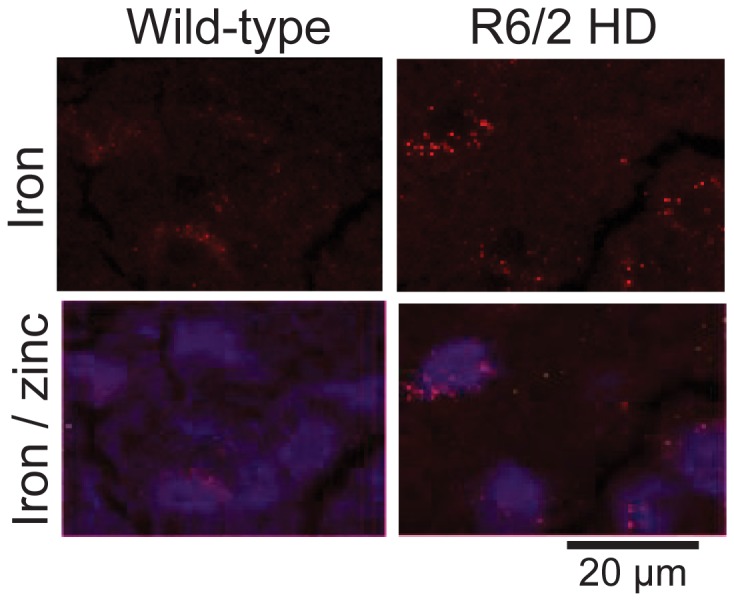
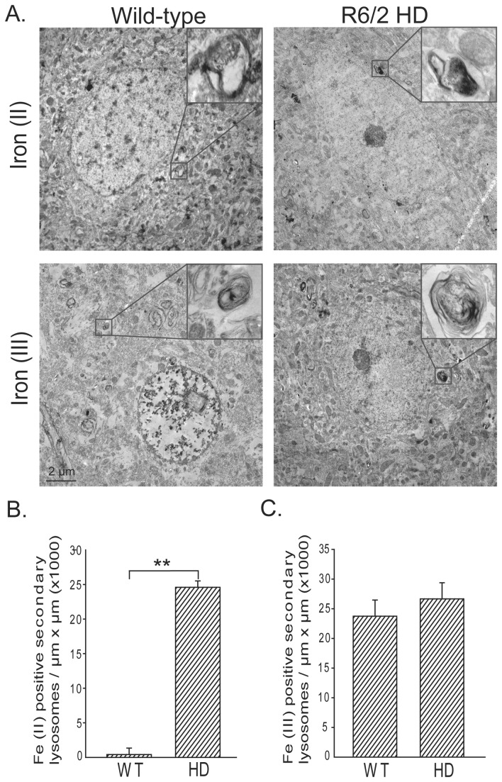
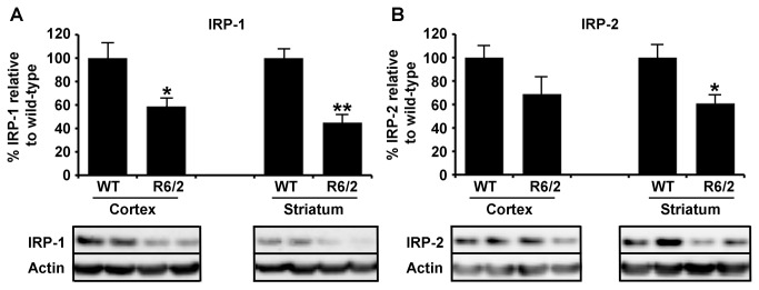
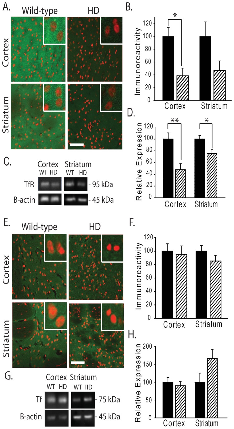
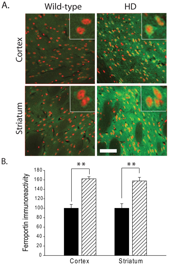
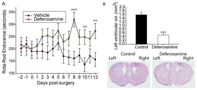
Huntington's disease (HD) is a progressive neurodegenerative disorder caused by a polyglutamine-encoding CAG expansion in the huntingtin gene. Iron accumulates in the brains of HD patients and mouse disease models. However, the cellular and subcellular sites of iron accumulation, as well as significance to disease progression are not well understood. We used independent approaches to investigate the location of brain iron accumulation. In R6/2 HD mouse brain, synchotron x-ray fluorescence analysis revealed iron accumulation as discrete puncta in the perinuclear cytoplasm of striatal neurons. Further, perfusion Turnbull's staining for ferrous iron (II) combined with transmission electron microscope ultra-structural analysis revealed increased staining in membrane bound peri-nuclear vesicles in R6/2 HD striatal neurons. Analysis of iron homeostatic proteins in R6/2 HD mice revealed decreased levels of the iron response proteins (IRPs 1 and 2) and accordingly decreased expression of iron uptake transferrin receptor (TfR) and increased levels of neuronal iron export protein ferroportin (FPN). Finally, we show that intra-ventricular delivery of the iron chelator deferoxamine results in an improvement of the motor phenotype in R6/2 HD mice. Our data supports accumulation of redox-active ferrous iron in the endocytic / lysosomal compartment in mouse HD neurons. Expression changes of IRPs, TfR and FPN are consistent with a compensatory response to an increased intra-neuronal labile iron pool leading to increased susceptibility to iron-associated oxidative stress. These findings, together with protection by deferoxamine, support a potentiating role of neuronal iron accumulation in HD.


Conflict of interest statement

Competing Interests: Co-authors Ashley I. Bush and James A. Duce are PLOS ONE Editorial Board members. Ashley Bush has shares in Prana Biotechnology Ltd, Mesoblast Ltd and Cogstate Ltd. This does not alter their adherence to all the PLOS ONE policies on sharing data and materials.

Figures

Figure 1
Figure 1. Iron accumulates in R6/2 HD striatal neurons.
Striata of 12-week-old R6/2 HD and wild-type litter mate mice were studied by synchrotron x-ray fluorescence (XRF - see methods). XRF identifies neuronal nuclei due to high zinc content (blue) [33]. HD mice demonstrate punctate peri-nuclear (cytoplasmic) accumulations of iron. Shown are representative images. n=2.
Figure 2
Figure 2. Iron (II) accumulates in R6/2 HD striatal neurons.
Iron (II) and iron (III) were determined by a modification of the perfusion Turnbull’s and Perl’s iron stains (see methods). A. Electron photomicrographs show striatal neurons with foci of iron (II) or (III) staining in membrane-bound structures consistent with secondary lysosomes. Quantification of iron (II) (B) and iron (III) (C) staining reveals significantly elevated iron (II) while iron (III) is unaltered. P-value: **< 0.01, n=2 and 5 neurons / mouse .
Figure 3
Figure 3. Decreased expression of iron-response proteins IRP1 and IRP2 in R6/2 HD mice.
A. IRP-1 protein expression in cortical and striatal separated brain tissue was significantly decreased in 12-week-old R6/2 mice compared to age-matched wild-type littermate controls. B. To a lesser extent IRP-2 expression was also decreased in HD model tissue from the same mice but did not reach significance in cortical regions. P-values: *<0.05, **< 0.01, n=5.
Figure 4
Figure 4. Decreased expression of transferrin receptor (TfR) but not transferrin (Tf) in R6/2 HD mice.
Analysis of TfR (A-D) and Tf (E-H) protein expression in brain. A. Representative images indicate decreased TfR immunoreactivity in HD TG cortex and striatum as compared to wild-type mice at 12-weeks of age. (Red, DRAQ5 stain; Green, TfR). Bar = 50 μm. Inserts = 20 μm. B. Quantification of neuronal cell body TfR immunoreactivity in cortex and striatum of wild-type and R6/2 mice. C-D. Western blot analysis of TfR detects a band migrating at ~95 kDa consistent with TfR (C); this band is significantly decreased in HD as compared to wild-type mice in cortex and striatum (D). E. Representative images of Tf immunofluorescence in HD and wild-type litter-mate mice. F. Quantification of Tf immunoreactivity. G-H. Western blot analysis of Tf detects a band migrating at ~75 kDa consistent with Tf (G); there is no change in protein levels in HD mice (H). P-values: *<0.05, **< 0.01, n=3-4 for immunofluorescence analyses and n=9-10 for Western blot analyses.
Figure 5
Figure 5. Increased expression of ferroportin (FPN) in R6/2 HD mice.
A. FPN immunoreactivity is increased in HD mouse cortical and striatal neurons as compared to wild-type mice at 12 weeks (red, DRAQ5 stain; green, FPN). Bar = 50 μm. Inserts = 20 μm. B. Quantification of A. FPN immunoreactivity is increased ~0.6 fold in both cortex and striatum of HD mice as compared to WT litter mate mice. P-value: **< 0.01, n=3-4.
Figure 6
Figure 6. Intra-ventricular delivery of the iron chelator deferoxamine provides protection in R6/2 HD mice.
Deferoxamine was delivered into the left ventricle of 6-week-old R6/2 HD mice at a dose-rate of 1-nmole / hour. Mice were analyzed daily before and after surgery for Rota-rod performance. A. Deferoxamine treated mice improve on Rota-Rod performance while vehicle-treated mice decline. B. Ventricular volumes are significantly lower in deferoxamine mice on the treated-side. P-values: **<0.01, ***<0.001: n=7.

Similar articles

Cited by

References

    1. the Huntington's Disease Collaborative Research Group (1993) A novel gene containing a trinucleotide repeat that is expanded and unstable on Huntington's disease chromosomes. Cell 72: 971-983. doi:10.1016/0092-8674(93)90585-E. PubMed: 8458085. - DOI - PubMed
    1. Pringsheim T, Wiltshire K, Day L, Dykeman J, Steeves T et al. (2012) The incidence and prevalence of Huntington's disease: a systematic review and meta-analysis. Mov Disord 27: 1083-1091. doi:10.1002/mds.25075. PubMed: 22692795. - DOI - PubMed
    1. Tabrizi SJ, Scahill RI, Durr A, Roos RA, Leavitt BR et al. (2011) Biological and clinical changes in premanifest and early stage Huntington's disease in the TRACK-HD study: the 12-month longitudinal analysis. Lancet Neurol 10: 31-42. doi:10.1016/S1474-4422(10)70276-3. PubMed: 21130037. - DOI - PubMed
    1. Browne SE, Beal MF (1994) Oxidative damage and mitochondrial dysfunction in neurodegenerative diseases. Biochem Soc Trans 22: 1002-1006. PubMed: 7698395. - PubMed
    1. Cui L, Jeong H, Borovecki F, Parkhurst CN, Tanese N et al. (2006) Transcriptional repression of PGC-1alpha by mutant huntingtin leads to mitochondrial dysfunction and neurodegeneration. Cell 127: 59-69. doi:10.1016/j.cell.2006.09.015. PubMed: 17018277. - DOI - PubMed

Publication types

MeSH terms