. 2013 Oct 16;8(10):e77751.
doi: 10.1371/journal.pone.0077751. eCollection 2013.

Enhanced production, purification, characterization and mechanism of action of salivaricin 9 lantibiotic produced by Streptococcus salivarius NU10

Affiliations

Enhanced production, purification, characterization and mechanism of action of salivaricin 9 lantibiotic produced by Streptococcus salivarius NU10

Abdelahhad Barbour et al. PLoS One. .

Abstract

Background: Lantibiotics are small lanthionine-containing bacteriocins produced by lactic acid bacteria. Salivaricin 9 is a newly discovered lantibiotic produced by Streptococcus salivarius. In this study we present the mechanism of action of salivaricin 9 and some of its properties. Also we developed new methods to produce and purify the lantibiotic from strain NU10.

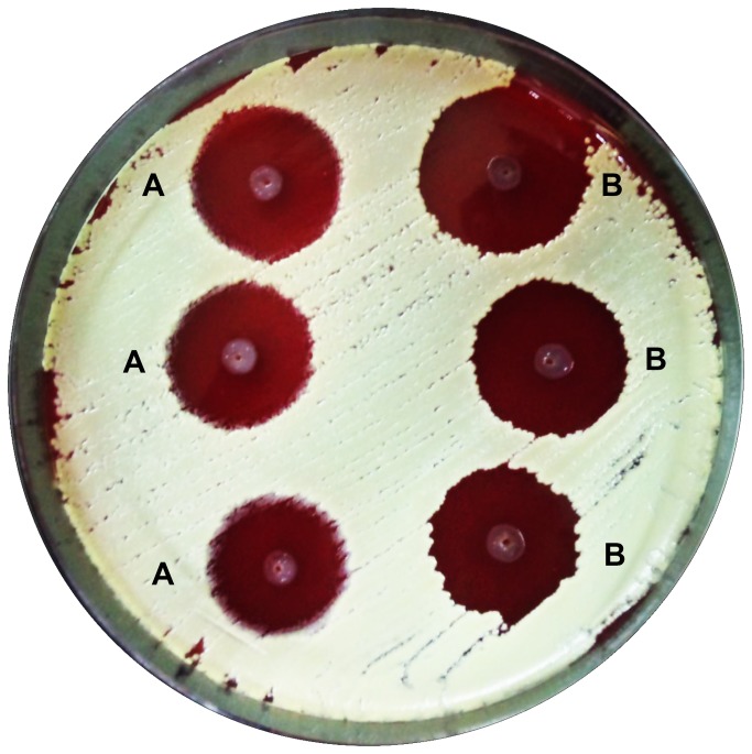
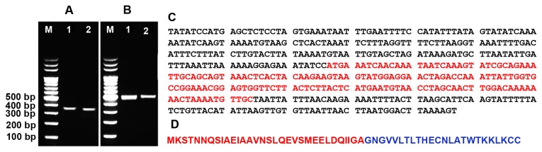
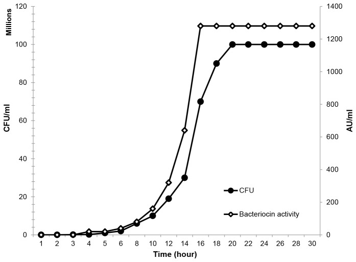
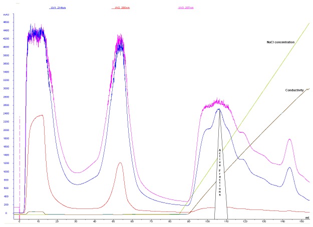
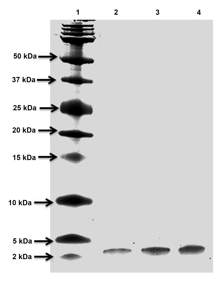
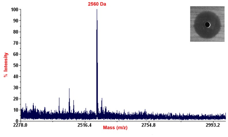
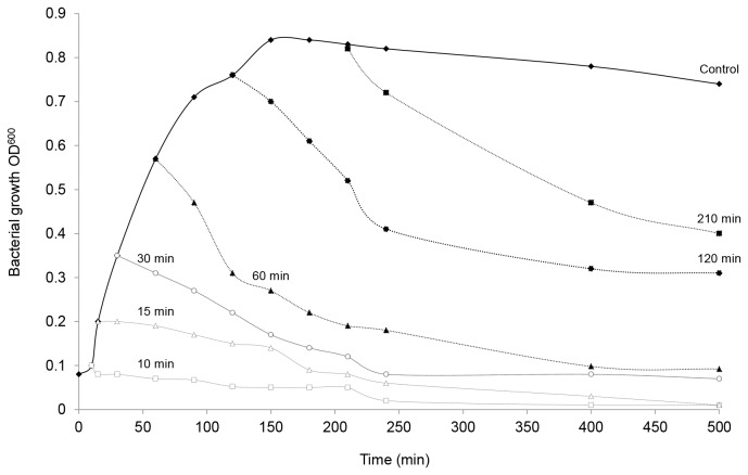
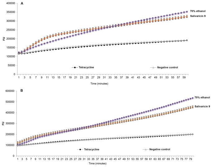
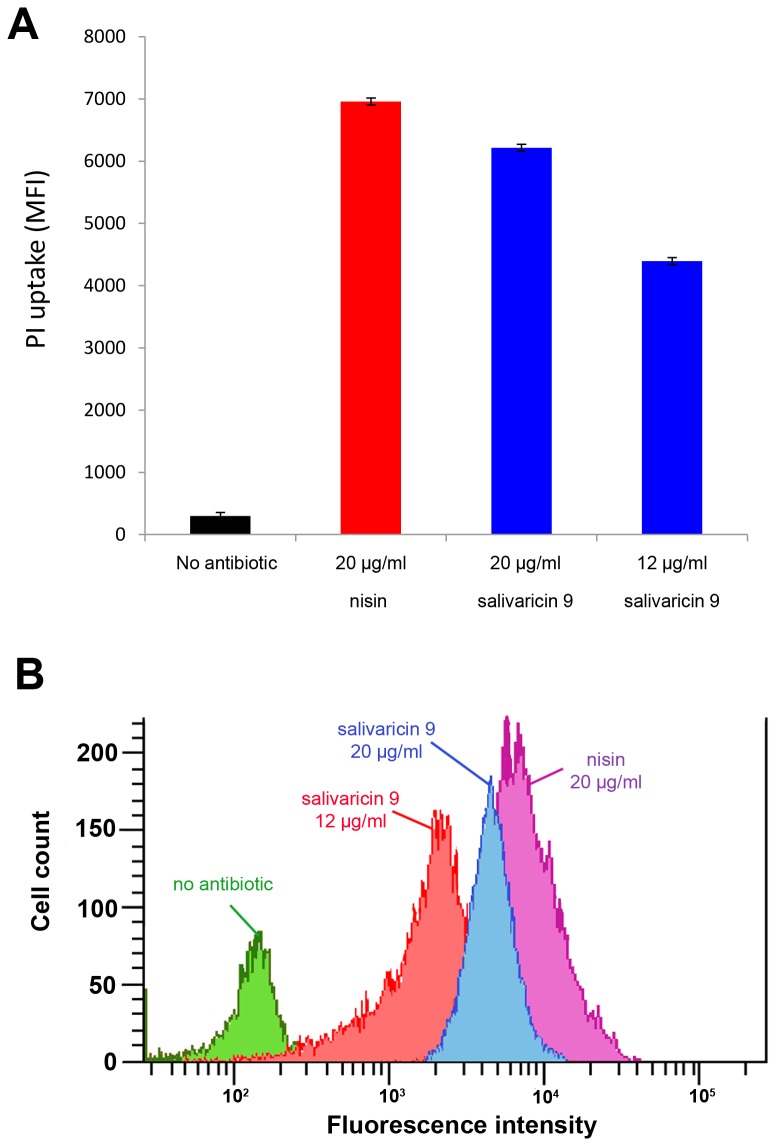
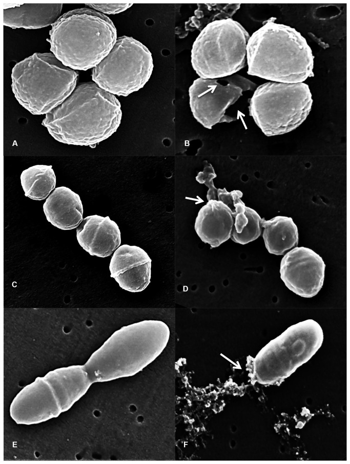
Methodology/principal findings: Salivaricin 9 was found to be auto-regulated when an induction assay was applied and this finding was used to develop a successful salivaricin 9 production system in liquid medium. A combination of XAD-16 and cation exchange chromatography was used to purify the secondary metabolite which was shown to have a molecular weight of approximately 3000 Da by SDS-PAGE. MALDI-TOF MS analysis indicated the presence of salivaricin 9, a 2560 Da lantibiotic. Salivaricin 9 is a bactericidal molecule targeting the cytoplasmic membrane of sensitive cells. The membrane permeabilization assay showed that salivaricin 9 penetrated the cytoplasmic membrane and induced pore formation which resulted in cell death. The morphological changes of test bacterial strains incubated with salivaricin 9 were visualized using Scanning Electron Microscopy which confirmed a pore forming mechanism of inhibition. Salivaricin 9 retained biological stability when exposed to high temperature (90-100°C) and stayed bioactive at pH ranging 2 to 10. When treated with proteinase K or peptidase, salivaricin 9 lost all antimicrobial activity, while it remained active when treated with lyticase, catalase and certain detergents.

Conclusion: The mechanism of antimicrobial action of a newly discovered lantibiotic salivaricin 9 was elucidated in this study. Salivaricin 9 penetrated the cytoplasmic membrane of its targeted cells and induced pore formation. This project has given new insights on lantibiotic peptides produced by S. salivarius isolated from the oral cavities of Malaysian subjects.


Conflict of interest statement

Competing Interests: The authors have declared that no competing interests exist.

Figures

Figure 1
Figure 1. Simultaneous Antagonism Assay.
A: NU10 was used as a bacteriocin producer, B: K12 (producer of salivaricin A and salivaricin B) was used as a positive control. Micrococcus luteus was used as target indicator strain.
Figure 2
Figure 2. Gene encoding salivaricin 9 production.
A: salA structural gene encoding salivaricin A production in strains NU10 (1) and YU10 (2). B: sivA structural gene encoding salivaricin 9 production in strains NU10 (1) and YU10 (2). (M) 100 bp DNA leader. Gel electrophoresis was performed using 2% (w/v) agarose and stained using GelRedTM. C: Assembled sivA gene sequence. The open reading frame ORF encoding the production of the leader and mature peptide is highlighted in red. D: In silico DNA to protein translation, leader peptide (red) and mature salivaricin 9 (blue).
Figure 3
Figure 3. Growth kinetics of strain NU10 during salivaricin 9 production.
Inhibitory activity of the cell free supernatant tested against Micrococcus luteus. Salivaricin 9 production was stable and consistent when strain NU10 reached the stationary phase of growth.
Figure 4
Figure 4. FPLC profile showing purification of salivaricin 9 using SP FF column.
Salivaricin 9 was bound to the strong cation exchanger efficiently and eluted using linear gradient of increasing NaCl concentrations. Salivaricin 9 was detected only at wave lengths of 207 and 214 nm.
Figure 5
Figure 5. Tris-Tricine SDS page of the purified peptide.
Lane 1: Dual Xtra protein marker (Bio Rad). Lanes: 2, 3 and 4: active fractions eluted from FPLC system.
Figure 6
Figure 6. MALDI-TOF MS analysis of salivaricin 9.
Active peak indicating the molecular weight of salivaricin 9 at 2560 Daltons.
Figure 7
Figure 7. Bactericidal mode of action of salivaricin 9.
Salivaricin 9 was added to different phases of bacterial growth. Salivaricin 9 induced bacterial lysis and decreased the indicator bacterial growth significantly. The sensitive bacteria Micrococcus luteus lost the ability to grow again after salivaricin 9 was added.
Figure 8
Figure 8. Membrane permeabilization assay of salivaricin 9.
A: Salivaricin 9 permeabilization activity towards cytoplasmic membrane of S. equisimilis. B: Salivaricin 9 permeabilization activity towards cytoplasmic membrane of Corynebacterium spp. Negative controls comprise targeted bacteria without adding salivaricin 9. Positive control used 70% ethanol. Tetracycline did not show any permeability activity in this test.
Figure 9
Figure 9. Flow cytometry analysis of pore-forming activity of salivaricin 9.
Like nisin, salivaricin 9 alters the membrane permeability of Micrococcus luteus ATCC10240 as measured by propidium iodide (PI) uptake. (A) Average MFI of triplicate measurements for nisin at a concentration of 20µg/ml and a range of salivaricin 9 concentrations of 3-fold and 5-fold above its MIC value. (B) Representative histogram of cell count versus PI fluorescence intensity at antibiotic concentrations shown in panel A.
Figure 10
Figure 10. morphological changes of sensitive bacterial cells incubated with salivaricin 9.
A: Untreated Micrococcus luteus used as a control. B: Morphological changes of Micrococcus luteus treated with salivaricin 9. C: Untreated S. equisimilis used as a control. D: Morphological changes of S. equisimilis treated with salivaricin 9. E: Untreated Corynebacterium spp used as a control. F: Morphological changes of Corynebacterium spp treated with salivaricin 9. White arrows indicate pores formed by salivaricin 9.

References

    1. Tiwari SK, Srivastava S (2008) Purification and characterization of plantaricin LR14: a novel bacteriocin produced by Lactobacillus plantarum LR/14. Appl Microbiol Biotechnol 79: 759-767. doi:10.1007/s00253-008-1482-6. PubMed: 18496687. - DOI - PubMed
    1. Klocke M, Mundt K, Idler F, Jung S, Backhausen JE (2005) Heterologous expression of enterocin A, a bacteriocin from Enterococcus faecium, fused to a cellulose-binding domain in Escherichia coli results in a functional protein with inhibitory activity against Listeria . Appl Microbiol Biotechnol 67: 532-538. doi:10.1007/s00253-004-1838-5. PubMed: 15660219. - DOI - PubMed
    1. Wan X, Li R, Saris PE, Takala TM (2012) Genetic characterisation and heterologous expression of leucocin C, a class IIa bacteriocin from Leuconostoc carnosum 4010. Appl Microbiol Biotechnol 97: 3509-3518. PubMed: 23053070. - PubMed
    1. Papagianni M, Anastasiadou S (2009) Pediocins: The bacteriocins of Pediococci. Sources, production, properties and applications. Microb Cell Factories 8: 1-16. doi:10.1186/1475-2859-8-1. - DOI - PMC - PubMed
    1. Abee T, Krockel L, Hill C (1995) Bacteriocins: modes of action and potentials in food preservation and control of food poisoning. Int J Food Microbiol 28: 169-185. doi:10.1016/0168-1605(95)00055-0. PubMed: 8750665. - DOI - PubMed

Publication types

MeSH terms