. 2013 Oct 16;8(10):e78050.
doi: 10.1371/journal.pone.0078050. eCollection 2013.

Salicylate prevents virus-induced type 1 diabetes in the BBDR rat

Affiliations

Salicylate prevents virus-induced type 1 diabetes in the BBDR rat

Chaoxing Yang et al. PLoS One. .

Abstract

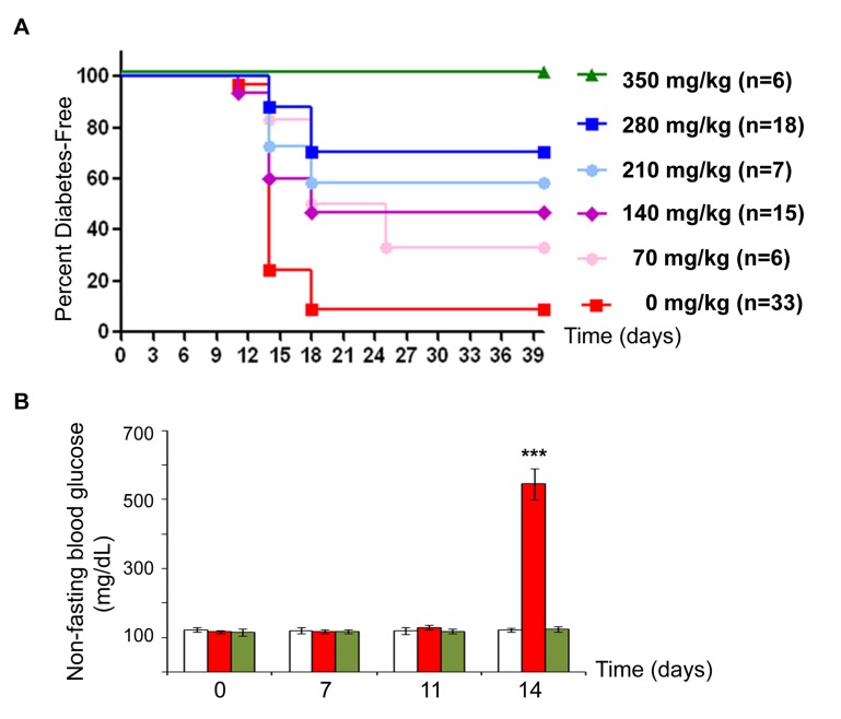
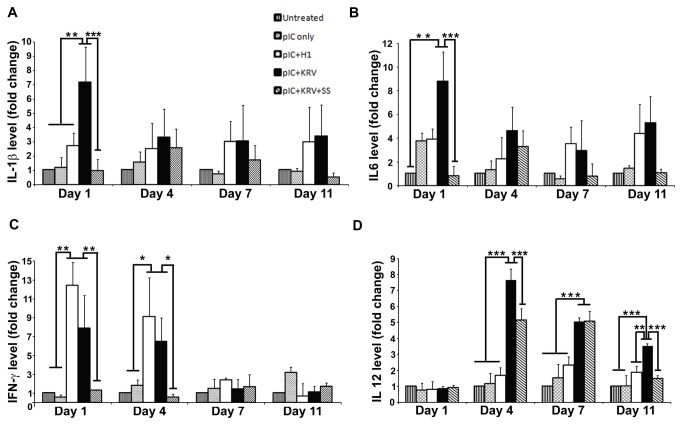
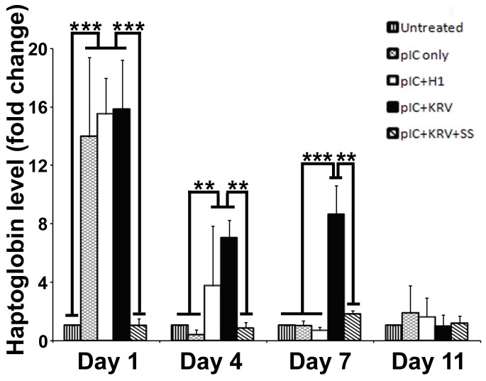
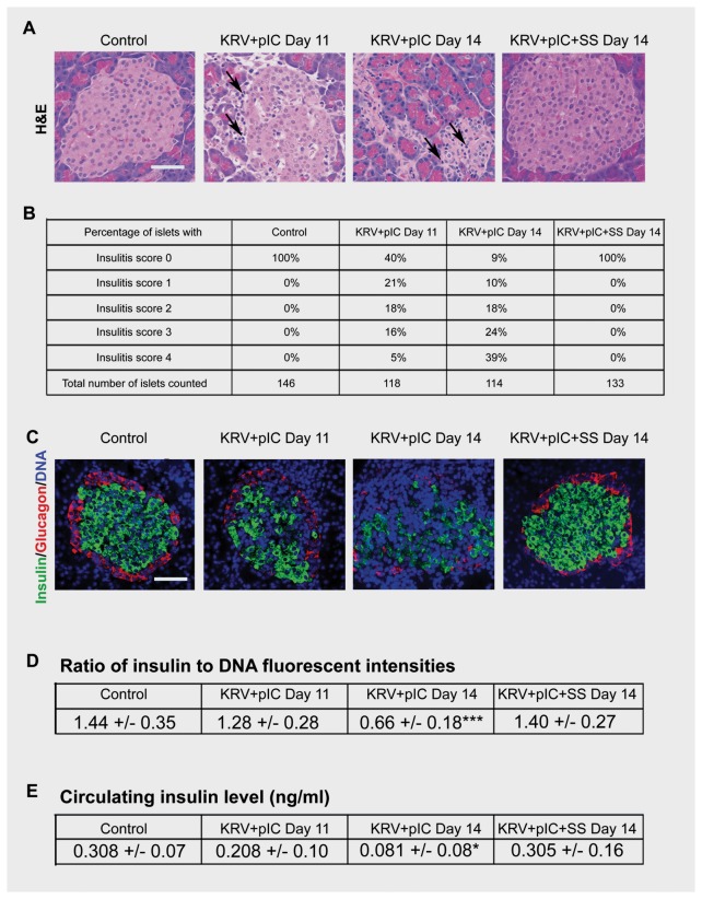
Epidemiologic and clinical evidence suggests that virus infection plays an important role in human type 1 diabetes pathogenesis. We used the virus-inducible BioBreeding Diabetes Resistant (BBDR) rat to investigate the ability of sodium salicylate, a non-steroidal anti-inflammatory drug (NSAID), to modulate development of type 1 diabetes. BBDR rats treated with Kilham rat virus (KRV) and polyinosinic:polycytidylic acid (pIC, a TLR3 agonist) develop diabetes at nearly 100% incidence by ~2 weeks. We found distinct temporal profiles of the proinflammatory serum cytokines, IL-1β, IL-6, IFN-γ, IL-12, and haptoglobin (an acute phase protein) in KRV+pIC treated rats. Significant elevations of IL-1β and IL-12, coupled with sustained elevations of haptoglobin, were specific to KRV+pIC and not found in rats co-treated with pIC and H1, a non-diabetogenic virus. Salicylate administered concurrently with KRV+pIC inhibited the elevations in IL-1β, IL-6, IFN-γ and haptoglobin almost completely, and reduced IL-12 levels significantly. Salicylate prevented diabetes in a dose-dependent manner, and diabetes-free animals had no evidence of insulitis. Our data support an important role for innate immunity in virus-induced type 1 diabetes pathogenesis. The ability of salicylate to prevent diabetes in this robust animal model demonstrates its potential use to prevent or attenuate human autoimmune diabetes.


Conflict of interest statement

Competing Interests: Dennis L. Guberski is President and C.E.O. and Christian W. Grant is a salaried employee of Biomedical Research Models. This does not alter their adherence to all the PLOS ONE policies on sharing data and materials.

Figures

Figure 1
Figure 1. Sodium salicylate improves blood glucose levels and diabetes-free survival of KRV+pIC treated rats.
BBDR rats were pre-treated with pIC and 0 to 350 mg/kg of sodium salicylate (SS) for 3 consecutive days (-3, -2, -1); KRV treatment and an additional treatment with SS was given on day 0. Doses of SS from 70 to 350 mg/kg body weight corresponded to 2- to 10-fold the amount given daily (for 14 weeks) to TINSAL-T2D patients [16]. (A) Kaplan-Meier curve showing onset of diabetes. As expected, >90% of rats given KRV+pIC without SS treatment became diabetic by 14-18 days, ***p<0.001. (B) Blood glucose levels were measured at the indicated times; n=6 for all groups, untreated (white), KRV+pIC (red), and KRV+pIC+SS (green); ***p<0.001, KRV+pIC vs. untreated and KRV+pIC+SS groups.
Figure 2
Figure 2. Proinfammatory cytokines are reduced in salicylate treated BBDR rats that remained diabetes-free.
BBDR rats were untreated (vertical), pre-treated with pIC alone (hatched), H1+pIC (clear), KRV+pIC (black), or KRV+pIC+SS (diagonal). Serum samples were collected from rats at 1, 4, 7, and 11 days following KRV or H1 treatment and analyzed for (A) IL-1β. (B) IL-6, (C) IFN-γ, and (D) IL-12. Untreated control, n=3; mean values of control rats for IL-1β. IL-6, IFN-γ, and IL-12 were 1.85, 0.54, 2.07, and 0.47 ng/ml, respectively. For all treated groups, n≥6. Mean and standard error are shown; *p<0.05; **p<0.01; ***p<0.001.
Figure 3
Figure 3. Salicylate blocks induction of the acute phase protein, haptoglobin.
BBDR rats were untreated (vertical), pre-treated with pIC alone (hatched), H1+pIC (clear), KRV+pIC (black), or KRV+pIC+SS (diagonal). Serum samples were collected from rats at 1, 4, 7, and 11 days following KRV treatment and analyzed for haptoglobin. Untreated control, n=3; all treated groups, n≥6. Mean and standard error are shown; **p<0.01; ***p<0.001.
Figure 4
Figure 4. Insulitis and subsequent loss of islet and serum insulin are completely abrogated in salicylate treated BBDR rats that remained diabetes-free.
BBDR rats were untreated or treated with KRV+pIC ± SS. (A) Pancreas samples were collected at the times indicated and stained for H&E (arrows indicate infiltrating lymphocytes). Panels shown are representative of n≥6 rats per group; scale bar, 50 µm. (B) Insulitis scores were determined from pancreas sections of 6-10 rats per group, 10-25 islets per rat. (C) Pancreas samples were collected and stained for insulin (green), glucagon (red), and DNA (DAPI, blue). Left panels are from an untreated (Control) BBDR rat, middle panels are from KRV+pIC treated rats at onset of insulitis (day 11) and hyperglycemia (day 14), and right panels are from a KRV+pIC+SS treated rat that was diabetes-free at day 14. Panels shown are representative of n≥3 rats per group; scale bar, 50 µm. (D) Insulin to DNA ratio is represented by green to blue intensity ratio; mean and standard error were calculated with n=9 islets per group, ***p<0.001, KRV+pIC vs. Control and KRV+pIC+SS groups. (E) Circulating serum levels of insulin were assayed at the indicated times; mean and standard error were calculated from n=6 rats per group, ***p<0.001, KRV+pIC vs. Control and KRV+pIC+SS groups.

References

    1. Castaño L, Eisenbarth GS (1990) Type-I diabetes: a chronic autoimmune disease of human, mouse, and rat. Annu Rev Immunol 8: 647-679. doi:10.1146/annurev.iy.08.040190.003243. PubMed: 2188676. - DOI - PubMed
    1. Redondo MJ, Yu L, Hawa M, Mackenzie T, Pyke DA et al. (2001) Heterogeneity of type I diabetes: analysis of monozygotic twins in Great Britain and the United States. Diabetologia 44: 354-362. doi:10.1007/s001250051626. PubMed: 11317668. - DOI - PubMed
    1. Nairn C, Galbraith DN, Taylor KW, Clements GB (1999) Enterovirus variants in the serum of children at the onset of Type 1 diabetes mellitus. Diabet Med 16: 509-513. doi:10.1046/j.1464-5491.1999.00098.x. PubMed: 10391400. - DOI - PubMed
    1. Yeung WC, Rawlinson WD, Craig ME (2011) Enterovirus infection and type 1 diabetes mellitus: systematic review and meta-analysis of observational molecular studies. BMJ 342: d35. doi:10.1136/bmj.d35. PubMed: 21292721. - DOI - PMC - PubMed
    1. Bortell R, Yang C (2012) The BB rat as a model of human type 1 diabetes. Methods Mol Biol 933: 31-44. PubMed: 22893399. - PubMed

Publication types