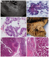
Duodenal subepithelial hyperechoic lesions of the third layer: Not always a lipoma
- PMID: 24147196
- PMCID: PMC3797905
- DOI: 10.4253/wjge.v5.i10.514
Duodenal subepithelial hyperechoic lesions of the third layer: Not always a lipoma
Abstract
Endoscopic ultrasonography is the most accurate procedure for the evaluation of subepithelial lesions. The finding of a homogeneous, hyperechoic, well-delimited lesion, originating from the third layer of the gastrointestinal tract (submucosa) suggests a benign tumor, generally lipoma. As other differential diagnoses have not been reported, echoendoscopists might not pursue a definitive pathological diagnosis or follow-up the patient. This case series aims to broaden the spectrum of differential diagnosis for duodenal hyperechoic third layer subepithelial lesions by providing four different and relevant pathologies with this echoendoscopic pattern.
Keywords: Duodenum; Endoscopic ultrasonography; Endoscopic ultrasound-guided fine needle aspiration; Lipoma; Subepithelial tumor.
Figures


Similar articles
-
Endoscopic ultrasonography diagnosis of subepithelial lesions.Dig Endosc. 2017 May;29(4):431-443. doi: 10.1111/den.12854. Epub 2017 Apr 6. Dig Endosc. 2017. PMID: 28258621 Review.
-
Subepithelial rectal gastrointestinal stromal tumor - the use of endoscopic ultrasound-guided fine needle aspiration to establish a definitive cytological diagnosis: a case report.J Med Case Rep. 2017 Mar 5;11(1):59. doi: 10.1186/s13256-017-1205-7. J Med Case Rep. 2017. PMID: 28259173 Free PMC article.
-
The role of endoscopic ultrasound in evaluation of gastric subepithelial lesions.Coll Antropol. 2010 Jun;34(2):757-62. Coll Antropol. 2010. PMID: 20698167 Review.
-
A case of gastric lipoma resected by endoscopic submucosa dissection with difficulty in preoperative diagnosis.Fukushima J Med Sci. 2017 Dec 19;63(3):160-164. doi: 10.5387/fms.2016-19. Epub 2017 Sep 14. Fukushima J Med Sci. 2017. PMID: 28904301 Free PMC article.
-
ENDOSCOPIC ULTRASOUND IN THE EVALUATION OF UPPER SUBEPITHELIAL LESIONS.Arq Gastroenterol. 2015 Jul-Sep;52(3):186-9. doi: 10.1590/S0004-28032015000300006. Arq Gastroenterol. 2015. PMID: 26486284
Cited by
-
Imaging of duodenal lipoma, case report of a rare tumor of the gastro intestinal tract.Radiol Case Rep. 2024 Oct 30;20(1):395-398. doi: 10.1016/j.radcr.2024.10.010. eCollection 2025 Jan. Radiol Case Rep. 2024. PMID: 39525895 Free PMC article.
-
Clinical Characteristics and Endoscopic Management of Duodenal Lipomas: 10-Year Experience from a Tertiary Center in China.Turk J Gastroenterol. 2023 Jul;34(7):720-727. doi: 10.5152/tjg.2023.22617. Turk J Gastroenterol. 2023. PMID: 37326152 Free PMC article.
References
-
- Rösch T, Lorenz R, Dancygier H, von Wickert A, Classen M. Endosonographic diagnosis of submucosal upper gastrointestinal tract tumors. Scand J Gastroenterol. 1992;27:1–8. - PubMed
-
- Săftoiu A, Vilmann P, Ciurea T. Utility of endoscopic ultrasound for the diagnosis and treatment of submucosal tumors of the upper gastrointestinal tract. Rom J Gastroenterol. 2003;12:215–229. - PubMed
-
- Taylor AJ, Stewart ET, Dodds WJ. Gastrointestinal lipomas: a radiologic and pathologic review. AJR Am J Roentgenol. 1990;155:1205–1210. - PubMed
LinkOut - more resources
Full Text Sources
Other Literature Sources
Research Materials

