. 2014 Mar 1;23(5):530-40.
doi: 10.1089/scd.2013.0297. Epub 2013 Dec 20.

Effects of long-term hypoxia and pro-survival cocktail in bone marrow-derived stromal cell survival

Affiliations

Effects of long-term hypoxia and pro-survival cocktail in bone marrow-derived stromal cell survival

Saejeong Kim et al. Stem Cells Dev. .

Abstract

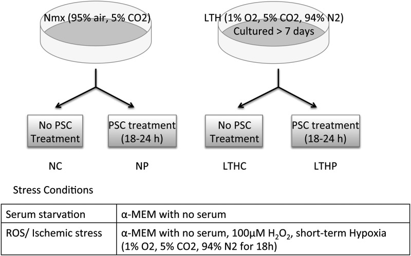
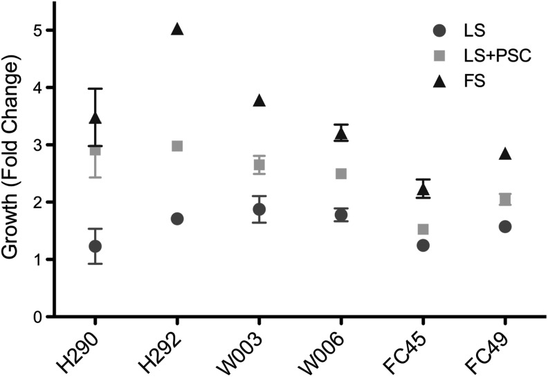
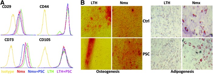
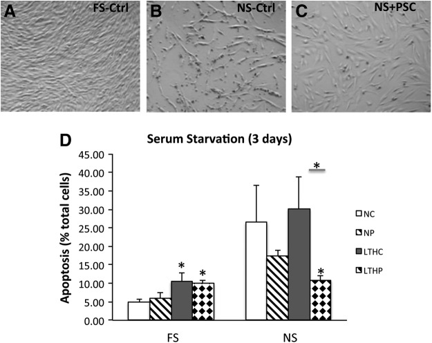
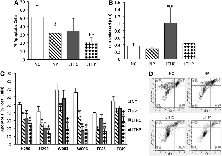
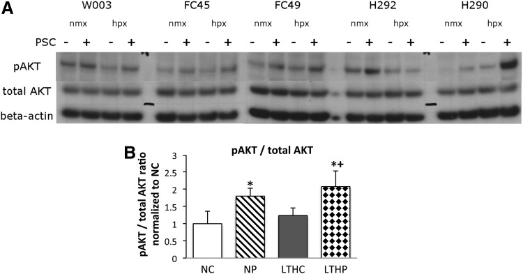
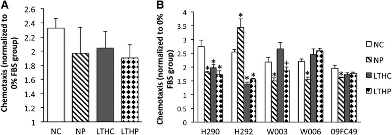
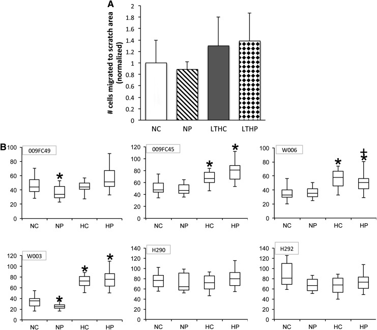
The goal of this study was to determine whether a pro-survival cocktail (PSC, consisting of IGF-1, Bcl-XL, and Caspase-I Inhibitor) and long-term hypoxia (LTH) enhance survival and functional properties of bone marrow-derived stromal stem cells (BMSCs), in response to stress conditions. PSC-treated cells retained BMSC surface markers and protected cells from apoptosis under serum starvation and ischemic (1% O2 and 100 μM H2O2) conditions. LTH promoted osteogenesis, while suppressing adipogenesis. LTH alone did not result in an improvement in the apoptosis rate; however, PSC conferred significant protection regardless of the oxygenation status. One of the possible mechanisms of PSC protection was due to the elevated phospho-AKT in treated groups. PSC treatment or LTH did not alter migration toward stem cell-derived factor-1 alpha (SDF-1α) or fetal bovine serum, nor did they enhance cell motility during wound healing. There was no difference in the secreted cytokine profiles of BMSCs treated with PSC after stress when grown in normoxic or LTH. However, LTH did upregulate the vascular endothelial growth factor, hepatocyte growth factor, and SDF-1α, while it downregulated other anti- and proinflammatory cytokines and chemokines. We also observed a high degree of interdonor BMSC variability in response to pretreatment with PSC and LTH, confounding the functional results, underscoring the observation that not all donor-derived BMSCs will respond similarly.


Figures

<b>FIG. 1.</b>
FIG. 1.
Graphic depiction of the preconditioning scheme and stress conditions.
<b>FIG. 2.</b>
FIG. 2.
Pro-survival cocktail (PSC)-treated cells maintain metabolic activity in low serum over 5 days. Cell proliferation and metabolic activity were measured using MTS assay in triplicates. Data shown is cell growth at 5 days.
<b>FIG. 3.</b>
FIG. 3.
PSC treatment did not alter phenotype and differentiation capability of bone marrow-derived stromal stem cells (BMSCs). (A) Representative histograms by fluorescence activated cell sorting (FACS) analysis of BMSC surface markers: CD29, CD44, CD73, and CD105. Long-term hypoxia (LTH) or PSC treatment did not change BMSC surface marker expression. The respective isotype controls are shown in a yellow line. (B) Osteogenic differentiation of BMSCs was confirmed by Alizarin Red staining (left panel); adipogenic differentiation of BMSCs was detected by Oil Red O staining (right panel). Original magnification was 20×.
<b>FIG. 4.</b>
FIG. 4.
PSC treatment protects BMSCs against serum starvation-induced apoptosis. (A) BMSCs grown in full serum. (B) BMSCs grown in no serum for 3 days showed blebbing and cell detachment. (C) PSC-treated cells maintain normal morphology after 3 days of serum deprivation. Original magnification was 20×. (D) Apoptosis was determined by flow cytometry after annexin V and PI staining on BMSCs cultured in FS or NS for 3 days before appropriate preconditioning regimen. FS, full serum; NS, no serum; NC, normoxia alone; NP, normoxia+PSC; LTHC, long-term hypoxia alone; LTHP, long-term hypoxia+PSC. *Indicates P<0.05 as compared with the NC group except otherwise indicated.
<b>FIG. 5.</b>
FIG. 5.
PSC treatment protects BMSCs against ROS and ischemia-induced apoptosis. (A) Apoptosis was determined by flow cytometry after annexin V and PI staining. Mean±SD of % apoptosis of six donor BMSCs. (B) LDH release was evaluated in the BMSC supernatant. Mean±SD of % apoptosis of six donor BMSCs. (C) Apoptosis in individual donor BMSCs showing donor variability. (D) Representative figures of flow cytometry after annexin V (y-axis) and PI (x-axis) staining are shown for donor W003 BMSCs. *Indicates P<0.05 and ** indicates P<0.01 as compared with the NC group. +Indicates P<0.05 compared with the LTHC group.
<b>FIG. 6.</b>
FIG. 6.
Protection conferred by PSC preconditioning is, in part, due to elevated phospho-Akt levels. Western blots were obtained from BMSCs cultured in long-term hypoxia or normoxia and pretreated with or without PSC. (A) Representative blots of phospho-Akt (pAkt), total Akt, and beta-actin are shown in different preconditioning groups. (B) Quantitative analysis of pAkt is shown using data obtained from three different blots. Data shown are mean±SD. *Indicates P<0.05 as compared with the NC group. +Indicates P<0.05 compared with the LTHC group.
<b>FIG. 7.</b>
FIG. 7.
LTH or PSC treatment had no significant effect on the migratory capacity of BMSCs. Using 10% FBS as a chemotactic agent in a modified Boyden chamber, the migration of BMSCs from the upper chamber across the membrane toward the lower chamber was measured overnight by CyQUANT fluorescence. The cell migration was normalized to the control with no chemotactic agent. (A) Average±SD of five donor-combined and normalized cell migration. (B) Chemotactic responses in five different donors after appropriate preconditioning show donor variability. *Indicates P<0.05 as compared with the NC group. +Indicates P<0.05 compared with the LTHC group.
<b>FIG. 8.</b>
FIG. 8.
Long-term hypoxia or PSC had no effect on cell motility toward wounds. The monolayers of BMSCs grown in tissue culture plates were scratched with a pipette tip to create wound. The locations were the migration will be measured was marked with a fine-tip marker, and then photomicrographs were taken at time 0 and 24 h to count cells migrated into the scratched area. (A) Quantitation of migrated cells toward wounds. Average±SD of six donor-combined and normalized cell migration. (B) Cell motility toward wounds in response to different preconditioning in individual donors. Number of cells migrated toward wounds. *Indicates P<0.05 as compared with the NC group. +Indicates P<0.05 compared with the HC group.

Similar articles

Cited by

References

    1. Lee JS, Hong JM, Moon GJ, Lee PH, Ahn YH. and Bang OY. (2010). A Long-Term Follow-Up Study of Intravenous Autologous Mesenchymal Stem Cell Transplantation in Patients With Ischemic Stroke. Stem Cells (Dayton, Ohio) 28:1099–1106 - PubMed
    1. Dharmasaroja P. (2009). Bone marrow-derived mesenchymal stem cells for the treatment of ischemic stroke. J Clin Neurosci 16:12–20 - PubMed
    1. Hu X, Yu SP, Fraser JL, Lu Z, Ogle ME, Wang J-A. and Wei L. (2008). Transplantation of hypoxia-preconditioned mesenchymal stem cells improves infarcted heart function via enhanced survival of implanted cells and angiogenesis. J Thorac Cardiovasc Surg 135:799–808 - PubMed
    1. Lee EJ, Park SJ, Kang SK, Kim G-H, Kang H-J, Lee S-W, Jeon HB. and Kim H-S. (2012). Spherical bullet formation via e-cadherin promotes therapeutic potency of mesenchymal stem cells derived from human umbilical cord blood for myocardial infarction. Mol Ther 20:1424–1433 - PMC - PubMed
    1. Yan J, Tie G, Xu TY, Cecchini K. and Messina LM. (2013). Mesenchymal stem cells as a treatment for peripheral arterial disease: current status and potential impact of type II diabetes on their therapeutic efficacy. Stem Cell Rev 9:360–372 - PMC - PubMed

Publication types

MeSH terms

LinkOut - more resources