. 2014 Mar;20(5-6):1089-95.
doi: 10.1089/ten.TEA.2013.0295. Epub 2013 Dec 11.

Neuronal electrophysiological function and control of neurite outgrowth on electrospun polymer nanofibers are cell type dependent

Affiliations

Neuronal electrophysiological function and control of neurite outgrowth on electrospun polymer nanofibers are cell type dependent

Justin L Bourke et al. Tissue Eng Part A. 2014 Mar.

Abstract

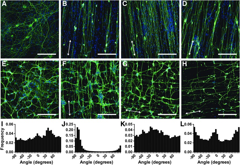
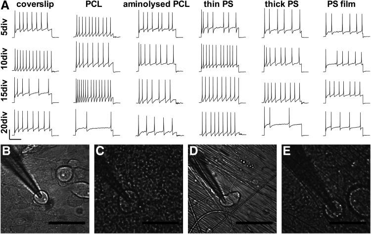
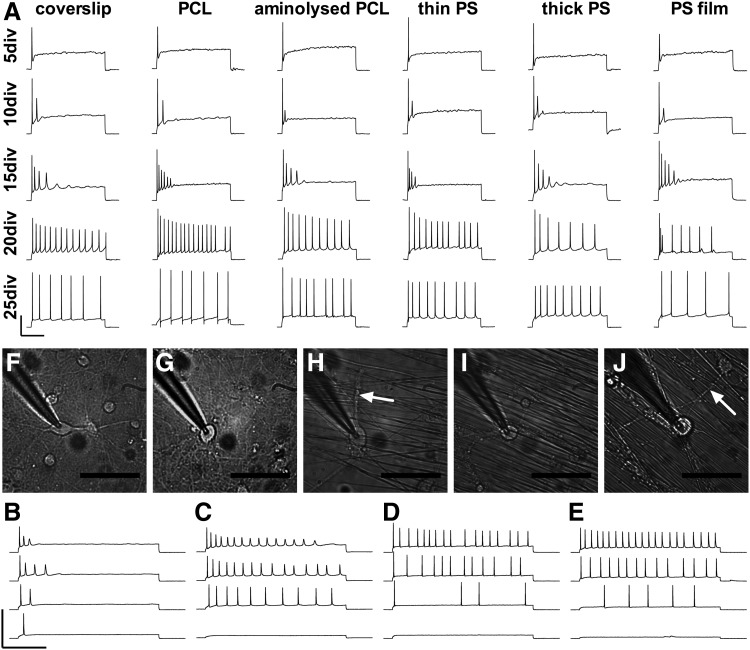
Modeling of cellular environments with nanofabricated biomaterial scaffolds has the potential to improve the growth and functional development of cultured cellular models, as well as assist in tissue engineering efforts. An understanding of how such substrates may alter cellular function is critical. Highly plastic central nervous system hippocampal cells and non-network forming peripheral nervous system dorsal root ganglion (DRG) cells from embryonic rats were cultured upon laminin-coated degradable polycaprolactone (PCL) and nondegradable polystyrene (PS) electrospun nanofibrous scaffolds with fiber diameters similar to those of neuronal processes. The two cell types displayed intrinsically different growth patterns on the nanofibrous scaffolds. Hippocampal neurites grew both parallel and perpendicular to the nanofibers, a property that would increase neurite-to-neurite contacts and maximize potential synapse development, essential for extensive network formation in a highly plastic cell type. In contrast, non-network-forming DRG neurons grew neurites exclusively along fibers, recapitulating the simple direct unbranching pathway between sensory ending and synapse in the spinal cord that occurs in vivo. In addition, the two primary neuronal types showed different functional capacities under patch clamp testing. The substrate composition did not alter the neuronal functional development, supporting electrospun PCL and PS as candidate materials for controlled cellular environments in culture and electrospun PCL for directed neurite outgrowth in tissue engineering applications.


Figures

<b>FIG. 1.</b>
FIG. 1.
TuJ1, a neuron-specific β-3 Tubulin antibody (green) and Dulbecco's modified Eagle's medium (nuclear, blue) immunostaining of dorsal root ganglion (DRG) (A–D) and hippocampal cells (E–H) at 5 days in vitro (DIV) on substrates of glass coverslips (A, E), aligned polycaprolactone (PCL) (B, F), aligned small diameter polystyrene (PS) scaffolds (C, G), and aligned large diameter PS scaffolds (D, H) (scale bar 200 μm). In each case, cells on glass coverslips sent out neurites in random orientation. While DRG cells sent out neurites along fibers, hippocampal cells sent out neurites both parallel and perpendicular to fibers. Direction histograms for DRG on coverslips (I) and thin PS fibers (J), and hippocampal cells on coverslips (K) and thin PS fibers (L) have been included. Arrows show the direction of fiber alignment.
<b>FIG. 2.</b>
FIG. 2.
(A) Embryonic day 18 (E18) rat DRG cells cultured on substrates and patch clamped at 5, 10, 15, and 20 DIV (scale 250 ms horizontal, 50 mV vertical). Cells on all substrates were capable of firing action potentials (APs) throughout the culture period in response to current injection. Optical micrographs of patched cells in the perfusion bath (scale bar 50 μm) on glass coverslips at 10 DIV (B) and 20 DIV (C), and PCL substrates at 10 DIV (D) and 20 DIV (E). Cultures at 20 days show a buildup of protein and lipid over the culture making patching of cells difficult. Electrophysiology on DRG neurons was from 4 independent cultures, with APs induced in 200 neurons up to 20 DIV.
<b>FIG. 3.</b>
FIG. 3.
(A) E18 rat hippocampal cells were cultured on substrates and patch clamped every 5 days out to 25 days in culture (scale 250 ms horizontal, 50 mV vertical). APs induced by current injection improved in function over time in culture, with no difference in functional development between substrates. Application of increasing current steps early in culture resulted in significant loss of amplitude of repeated APs (B: 15 DIV, C: 20 DIV), a trend that persisted until cell maturity at 25 days in culture (D). Function at 50 DIV (E) was similar to 25 DIV, suggesting cellular functional maturity from 25 days in culture (scale 250 ms horizontal, 200 mV vertical). Optical micrographs (scale 50 μm) of patched hippocampal cells on glass coverslips at 10 DIV (F) and 25 DIV (G) and PCL substrates at 10 DIV (H) and 25 DIV (I) show clean and consistent extended cultures, remaining clean out to 50 days in culture (J). Arrows point to hippocampal neurites clearly growing perpendicular to fibers. Electrophysiology on hippocampal neurons was from 5 independent cultures, with APs induced in 129 neurons up to 25 DIV and another 4 neurons at 50 DIV.

References

    1. Hutmacher D.W.Biomaterials offer cancer research the third dimension. Nat Mater 9,90, 2010 - PubMed
    1. Lutton C., and Goss B.Caring about microenvironments. Nat Biotechnol 26,613, 2008 - PubMed
    1. Edelman D.B., and Keefer E.W.A cultural renaissance: in vitro cell biology embraces three-dimensional context. Exp Neurol 192,1, 2005 - PubMed
    1. Kleinfeld D., Kahler K.H., and Hockberger P.E.Controlled outgrowth of dissociated neurons on patterned substrates. J Neurosci 8,4098, 1988 - PMC - PubMed
    1. Magnusson A.K., Linderholm P., Vieider C., Ulfendahl M., and Erlandsson A.Surface protein patterns govern morphology, proliferation, and expression of cellular markers but have no effect on physiological properties of cortical precursor cells. J Neurosci Res 86,2363, 2008 - PubMed

LinkOut - more resources