. 2014 Jan 17;9(1):156-63.
doi: 10.1021/cb400548s. Epub 2013 Nov 7.

Molecular grafting onto a stable framework yields novel cyclic peptides for the treatment of multiple sclerosis

Affiliations

Molecular grafting onto a stable framework yields novel cyclic peptides for the treatment of multiple sclerosis

Conan K Wang et al. ACS Chem Biol. .

Abstract

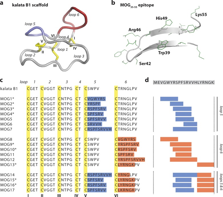
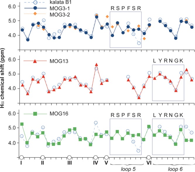
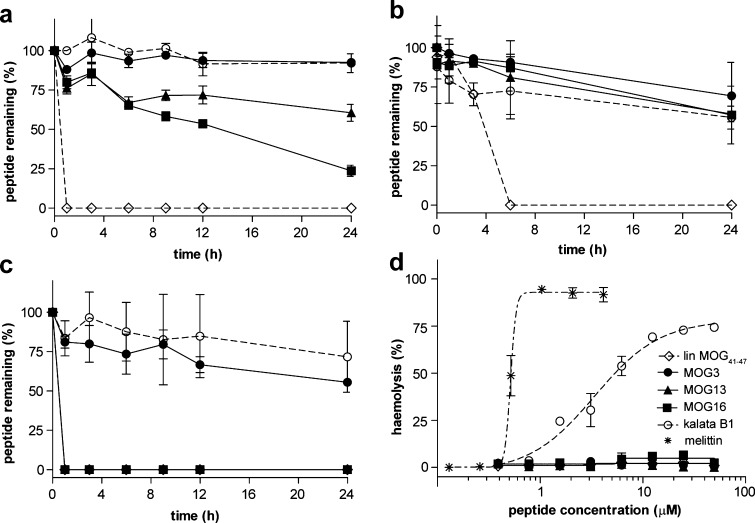
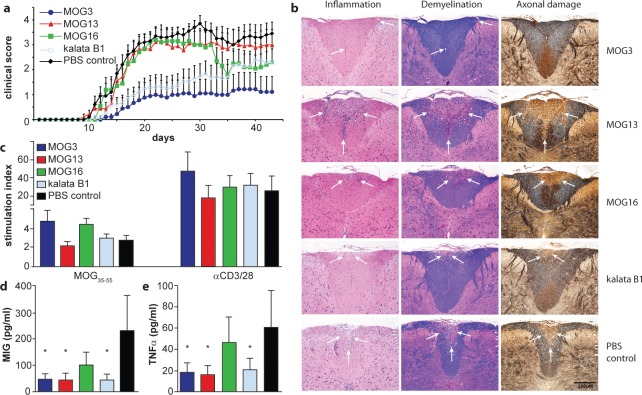
Multiple sclerosis (MS) is an inflammatory disease of the central nervous system (CNS) and is characterized by the destruction of myelin and axons leading to progressive disability. Peptide epitopes from CNS proteins, such as myelin oligodendrocyte glycoprotein (MOG), possess promising immunoregulatory potential for treating MS; however, their instability and poor bioavailability is a major impediment for their use clinically. To overcome this problem, we used molecular grafting to incorporate peptide sequences from the MOG35-55 epitope onto a cyclotide, which is a macrocyclic peptide scaffold that has been shown to be intrinsically stable. Using this approach, we designed novel cyclic peptides that retained the structure and stability of the parent scaffold. One of the grafted peptides, MOG3, displayed potent ability to prevent disease development in a mouse model of MS. These results demonstrate the potential of bioengineered cyclic peptides for the treatment of MS.


Figures

Figure 1
Figure 1
Molecular grafting of antigenic peptides onto a cyclotide scaffold. (a) The cyclotide kalata B1 is stabilized by three conserved disulfide bonds (shown in yellow) and a head-to-tail cyclized backbone, which together form the cyclic cystine knot motif. The six conserved cysteines (numbered with roman numerals) divide the backbone into six loops, including loops 5 and 6 that are amenable to molecular grafting and colored in blue and red, respectively. (b) Native structure of the MOG35–55 bioactive epitope extracted from the three-dimensional structure of the entire MOG (myelin oligodendrocyte glycoprotein) protein. The epitope comprises two antiparallel β-sheets; selected residues are numbered in single letter code, and amino acid side chains are shown in green. (c) Aligned sequences of kalata B1 and novel grafted molecules MOG1–17. The six cysteine residues are highlighted in yellow, and the six loops are numbered. The grafted sequences in loops 5 and 6 are highlighted in blue and red, respectively. Grafted MOG peptides that adopted a native-like globular fold are marked with an asterisk. (d) The MOG35–55 bioactive epitope was grafted into the kalata B1 scaffold by dividing the full epitope sequence (shown on top) into smaller fragments and inserting them into either loop 5, loop 6 or loops 5 and 6.
Figure 2
Figure 2
Structural characteristics of MOG-grafted peptides. The Hα chemical shifts of MOG-grafted peptides and kalata B1 are shown. The positions of cysteines are indicated on the horizontal axis to align the sequence (non-cysteine residues are not shown for clarity). Two conformations for MOG3 (referred to as MOG3-1 and MOG3-2 in this figure) were observed. The different symbols used for each peptide are shown. Segments of the parent scaffold that have been grafted onto in loops 5 and 6 are boxed, and the sequences of the grafted epitopes are also shown.
Figure 3
Figure 3
Stability and hemolytic activity of MOG-grafted peptides. Stability of grafted peptides and controls over time (0–24 h) in (a) human serum, (b) hydrochloric acid, and (c) pancreatin as measured by the percentage of remaining intact peptide. (d) Hemolytic activity of grafted peptides was measured by the propensity of lysing human red blood cells. Mellitin, a strong hemolytic peptide from bee venom, was used as control. A legend for all peptides is shown.
Figure 4
Figure 4
Activity of novel grafted peptides in vivo in experimental autoimmune encephalomyelitis in mice. (a) Clinical score of EAE mice after vaccination with MOG-grafted peptides (MOG3, dark blue line; MOG13, red line; MOG16, green line) and controls (kalata B1, light blue line; PBS, black line) was monitored. (b) The influence of MOG-grafted cyclotide vaccination on the formation of CNS inflammatory and demyelinating lesions was examined by histological studies of fixed tissue using hematoxylin/eosin, Luxol fast blue, and Bielshowsky silver staining. Regions of inflammation, demyelination, and axonal damage are highlighted by white arrows. (c) Proliferation of spleen cells in response to the encephalitogen MOG35–55 and stimulation by the polyclonal activators, anti-CD3 and anti-CD28 antibodies. (d, e) Significantly reduced levels of the chemokine MIG (d) and TNFα (e) were demonstrated in non-stimulated spleen cell supernatants generated from animals treated with MOG3, MOG13, and kalata B1. * p < 0.05 compared to PBS control.
Figure 5
Figure 5
Effect of grafted peptides on the proliferation of spleen cells from 2D2 TCR transgenic mice. Proliferation of spleen cells from 2D2 TCR transgenic mice stimulated with different concentrations of MOG35–55 and incubated with (a) kalata B1, (b) MOG3, (c) MOG13, and (d) MOG16. Results show the mean ± SEM of three independent experiments (n = 3). * p < 0.05, *** p < 0.001.

Similar articles

Cited by

References

    1. Ewing C.; Bernard C. C. (1998) Insights into the aetiology and pathogenesis of multiple sclerosis. Immunol. Cell Biol. 76, 47–54. - PubMed
    1. Bernard C. C.; Kerlero de Rosbo N. (1992) Multiple sclerosis: an autoimmune disease of multifactorial etiology. Curr. Opin. Immunol. 4, 760–765. - PubMed
    1. Onuki M.; Ayers M. M.; Bernard C. C.; Orian J. M. (2001) Axonal degeneration is an early pathological feature in autoimmune-mediated demyelination in mice. Microsc. Res. Tech. 52, 731–739. - PubMed
    1. Ayers M. M.; Hazelwood L. J.; Catmull D. V.; Wang D.; McKormack Q.; Bernard C. C.; Orian J. M. (2004) Early glial responses in murine models of multiple sclerosis. Neurochem. Int. 45, 409–419. - PubMed
    1. Bernard C. C.; Johns T. G.; Slavin A.; Ichikawa M.; Ewing C.; Liu J.; Bettadapura J. (1997) Myelin oligodendrocyte glycoprotein: a novel candidate autoantigen in multiple sclerosis. J. Mol. Med. 75, 77–88. - PubMed

Publication types

MeSH terms

Substances