

Blood-brain-barriers in aging and in Alzheimer's disease
- PMID: 24148264
- PMCID: PMC4015275
- DOI: 10.1186/1750-1326-8-38
Blood-brain-barriers in aging and in Alzheimer's disease
Abstract
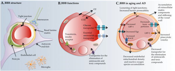
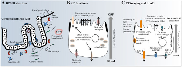
The aging process correlates with a progressive failure in the normal cellular and organ functioning; these alterations are aggravated in Alzheimer's disease (AD). In both aging and AD there is a general decrease in the capacity of the body to eliminate toxic compounds and, simultaneously, to supply the brain with relevant growth and nutritional factors. The barriers of the brain are targets of this age related dysfunction; both the endothelial cells of the blood-brain barrier and the choroid plexus epithelial cells of the blood-cerebrospinal fluid barrier decrease their secretory capacity towards the brain and their ability to remove toxic compounds from the brain. Additionally, during normal aging and in AD, the permeability of the brain barriers increase. As such, a greater contact of the brain parenchyma with the blood content alters the highly controlled neural environment, which impacts on neural function. Of interest, the brain barriers are more than mere obstacles to the passage of molecules and cells, and therefore active players in brain homeostasis, which is still to be further recognized and investigated in the context of health and disease. Herein, we provide a review on how the brain barriers change during aging and in AD and how these processes impact on brain function.
Figures
References
-
- Brookmeyer R, Johnson E, Ziegler-Graham K, Arrighi HM. Forecasting the global burden of Alzheimer’s disease. Alzheimers Dement. 2007;8:186–191. - PubMed
-
- Selkoe DJ. Alzheimer’s disease: genes, proteins, and therapy. Physiol Rev. 2001;8:741–766. - PubMed
-
- Michaud M, Balardy L, Moulis G, Gaudin C, Peyrot C, Vellas B, Cesari M, Nourhashemi F. Proinflammatory Cytokines, Aging, and Age-Related Diseases. J Am Med Dir Assoc. 2013;8(13):00280–00286. - PubMed
-
- Abbott NJ, Patabendige AA, Dolman DE, Yusof SR, Begley DJ. Structure and function of the blood–brain barrier. Neurobiol Dis. 2010;8:13–25. - PubMed
Publication types
MeSH terms
LinkOut - more resources
Full Text Sources
Other Literature Sources
Medical
Research Materials
