. 2013 Oct 22;13(1):103.
doi: 10.1186/1475-2867-13-103.

High expression of prolactin receptor is associated with cell survival in cervical cancer cells

Affiliations

High expression of prolactin receptor is associated with cell survival in cervical cancer cells

Edgar I Lopez-Pulido et al. Cancer Cell Int. .

Abstract

Background: The altered expression of prolactin (PRL) and its receptor (PRLR) has been implicated in breast and other types of cancer. There are few studies that have focused on the analysis of PRL/PRLR in cervical cancer where the development of neoplastic lesions is influenced by the variation of the hormonal status. The aim of this study was to evaluate the expression of PRL/PRLR and the effect of PRL treatment on cell proliferation and apoptosis in cervical cancer cell lines.

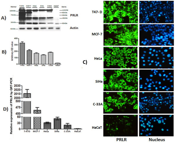
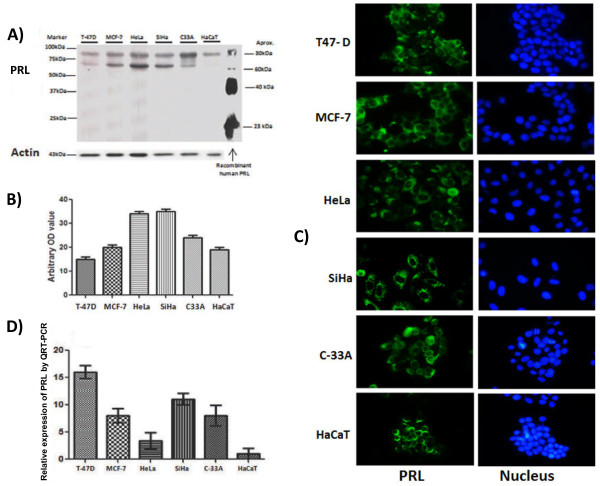
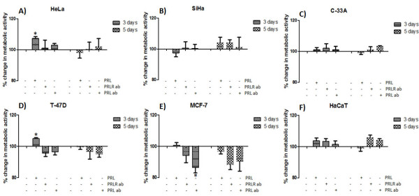
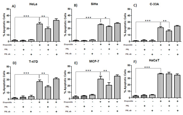
Results: High expression of multiple PRLR forms and PRLvariants of 60-80 kDa were observed in cervical cancer cell lines compared with non-tumorigenic keratinocytes evaluated by Western blot, immunofluorecence and real time PCR. Treatment with PRL (200 ng/ml) increased cell proliferation in HeLa cells determined by the MTT assay at day 3 and after 1 day a protective effect against etoposide induced apoptosis in HeLa, SiHa and C-33A cervical cancer cell lines analyzed by the TUNEL assay.

Conclusions: Our data suggests that PRL/PRLR signaling could act as an important survival factor for cervical cancer. The use of an effective PRL antagonist may provide a better therapeutic intervention in cervical cancer.


Figures

Figure 1
Figure 1
PRLR expression in human cervical cancer cells. SiHa, C-33A, HeLa (Cervical cancer cells) and control cells MCF-7, T-47D (breast cancer), HaCaT (Inmortalized human keratinocytes) were cultured in DMEM or RPMI medium containing 10% FBS. A) PRLR protein was determined by western blot using a specific antibody against the PRLR, PRLR proteins were identified by their size. B) Demonstration of the arbitrary optical density measurements from Western immunoblots assessing PRLR levels. C) The cells grown on coverslips were fixed, and the localization of PRLR (green) was observed by inmunocitochemistry using a secondary antibody conjugated with Alexa fluor 488 and DAPI stain (blue) to visualize the presence of cells. Magnification 10 x. D) Relative expression of PRLR mRNA was measure by quantitative RT-PCR.
Figure 2
Figure 2
Presence of autocrine PRL in human cervical cancer cell lines. SiHa, C-33A, HeLa (Cervical cancer cells) and control cells MCF-7, T-47D (breast cancer), HaCaT (Inmortalized human keratinocytes) were cultured in DMEM or RPMI medium containing 10% FBS. A) PRL protein was determined by western blot using a specific antibody against PRL. B) Demonstration of the arbitrary optical density measurements from Western immunoblots assessing PRL levels. C) The cells grown on coverslips were fixed, and the localization of PRL (green) was observed by inmunocitochemistry using a secondary antibody conjugated with Alexa fluor 488 and DAPI stain (blue) to visualize the presence of cells. Magnification 40 x. D) Relative expression of PRL mRNA was measure by quantitative RT-PCR.
Figure 3
Figure 3
Effects of PRL and PRL or PRLR blocking antibodies on proliferation of cervical cancer cells. Effects on metabolic activity after the incubation with PRL (200 ng/ml), PRL-AB (200 ng/ml) or PRLR-AB (2.5 μg) for 3 or 5 days in HeLa, SiHa, C-33A (A, B, C) and control cells MCF-7, T-47D and HaCaT (D, E, F). Graphs show experiments performed in triplicate, which are repeated at least three times. *p<.05.
Figure 4
Figure 4
Effects of PRL and PRL antibody on apoptosis induced by etoposide. Effects of the treatment with etoposide (40 ug/ml) for 24 hrs with or without added PRL (200 ng/ml) or PRL-AB (200 ng/ml) in SiHa, C-33 A, HeLa (A, B, C) and control cells MCF-7, T-47D and HaCaT (D, E, F). DNA strand breaks were analyzed microscopically by TUNEL. Graphs represent the mean of at least three replicate, where *p<0.05, **p<0.01 and ***p<.001.

Similar articles

Cited by

References

    1. Goffin V, Binart N, Touraine P, Kelly PA. Prolactin: the new biology of an old hormone. Annu Rev Physiol. 2002;13:47–67. - PubMed
    1. Welsch CW, Nagasawa H. Prolactin and murine mammary tumorigenesis: a review. Cancer Res. 1977;13(4):951–963. - PubMed
    1. Clevenger CV, Furth PA, Hankinson SE, Schuler LA. The role of prolactin in mammary carcinoma. Endocr Rev. 2003;13(1):1–27. - PMC - PubMed
    1. Tworoger SS, Hankinson SE. Prolactin and breast cancer etiology: an epidemiologic perspective. Journal J Mammary Gland Biol Neoplasia. 2008;13(1):41–53. - PubMed
    1. Bonneterre J, Mauriac L, Weber B, Roche H, Fargeot P, Tubiana-Hulin M, Sevin M, Chollet P, Cappelaere P. Tamoxifen plus bromocriptine versus tamoxifen plus placebo in advanced breast cancer: results of a double blind multicentre clinical trial. Eur J Cancer Clin Oncol. 1988;13(12):1851–1853. - PubMed

LinkOut - more resources