. 2014 Mar;146(3):776-88.
doi: 10.1053/j.gastro.2013.10.019. Epub 2013 Oct 19.

Extensive conversion of hepatic biliary epithelial cells to hepatocytes after near total loss of hepatocytes in zebrafish

Affiliations

Extensive conversion of hepatic biliary epithelial cells to hepatocytes after near total loss of hepatocytes in zebrafish

Tae-Young Choi et al. Gastroenterology. 2014 Mar.

Abstract

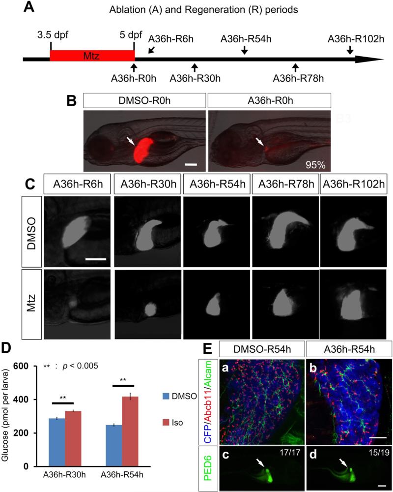
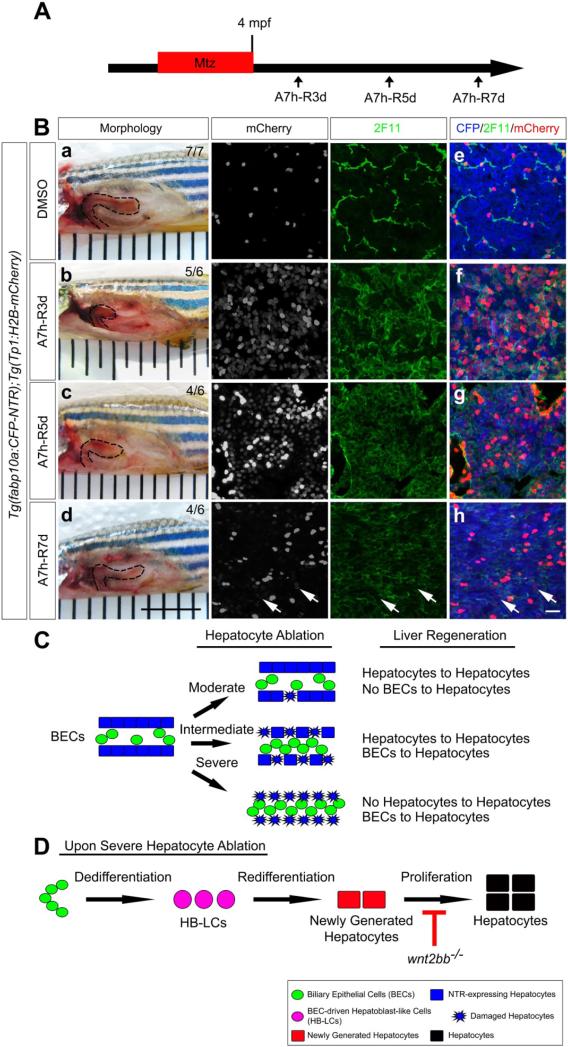
Background & aims: Biliary epithelial cells (BECs) are considered to be a source of regenerating hepatocytes when hepatocyte proliferation is compromised. However, there is still controversy about the extent to which BECs can contribute to the regenerating hepatocyte population, and thereby to liver recovery. To investigate this issue, we established a zebrafish model of liver regeneration in which the extent of hepatocyte ablation can be controlled.

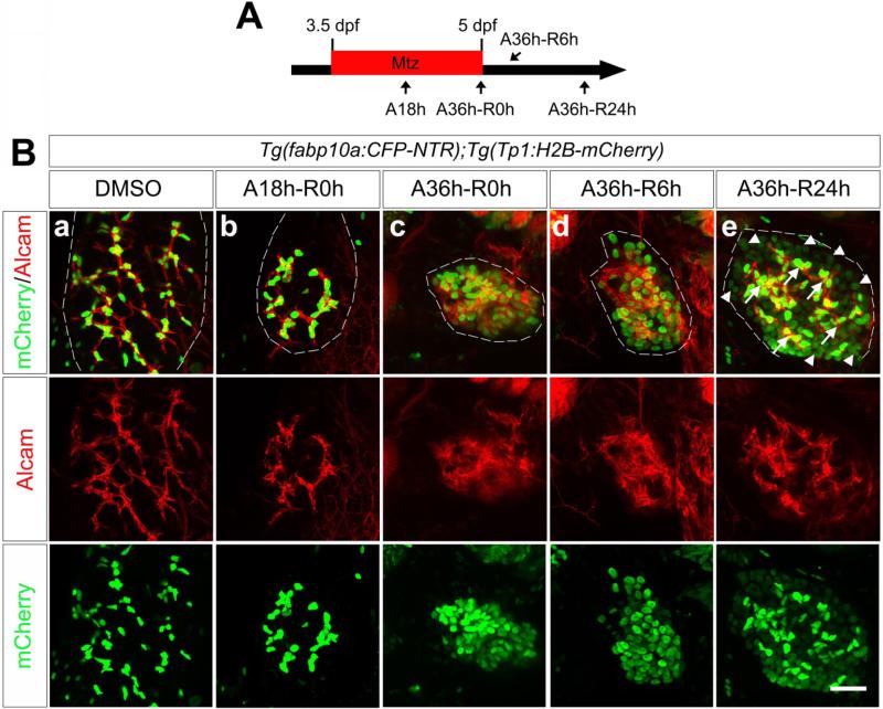
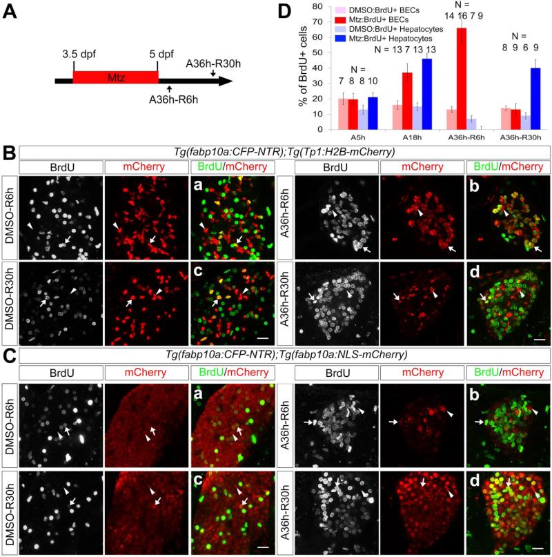
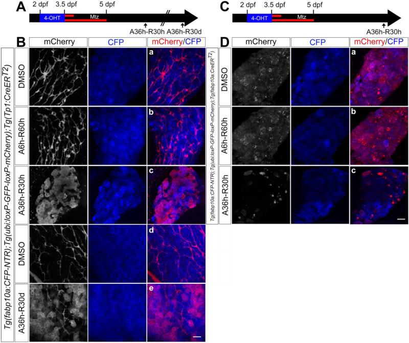
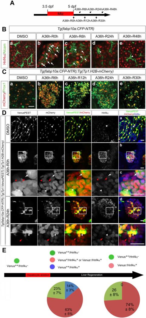
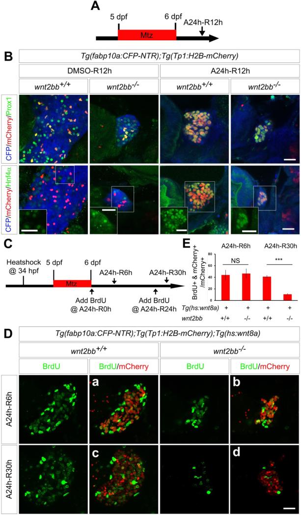
Methods: Hepatocytes were depleted by administration of metronidazole to Tg(fabp10a:CFP-NTR) animals. We traced the origin of regenerating hepatocytes using short-term lineage-tracing experiments, as well as the inducible Cre/loxP system; specifically, we utilized both a BEC tracer line Tg(Tp1:CreER(T2)) and a hepatocyte tracer line Tg(fabp10a:CreER(T2)). We also examined BEC and hepatocyte proliferation and liver marker gene expression during liver regeneration.

Results: BECs gave rise to most of the regenerating hepatocytes in larval and adult zebrafish after severe hepatocyte depletion. After hepatocyte loss, BECs proliferated as they dedifferentiated into hepatoblast-like cells; they subsequently differentiated into highly proliferative hepatocytes that restored the liver mass. This process was impaired in zebrafish wnt2bb mutants; in these animals, hepatocytes regenerated but their proliferation was greatly reduced.

Conclusions: BECs contribute to regenerating hepatocytes after substantial hepatocyte depletion in zebrafish, thereby leading to recovery from severe liver damage.

Keywords: Dedifferentiation; Liver Regeneration; Oval Cells; Stem Cells.


Figures

Figure 1
Figure 1
A zebrafish liver regeneration model. (A) Scheme illustrating the periods of Mtz treatment (A, ablation) and liver regeneration (R). (B, C) To reveal liver size, CFP expression from Tg(fabp10a:CFP-NTR) larvae (B, red; C, white) was examined right after hepatocyte ablation (B) and during liver regeneration (C) under an epifluorescence microscope. (D) Glucose levels in the regenerating larvae treated with DMSO or isoprenaline (Iso) for 30 hours after hepatocyte ablation. (E) Confocal images showing whole-mount immunostaining of the regenerating liver reveal the correct location of bile canaliculi between hepatocytes and BECs, as assessed by Abcb11 (red) and Alcam (green) expression (Ea-Eb). Epifluorescence images of regenerating larvae treated with PED6 reveal the accumulation of the processed PED6 in the gallbladder at A36h-R54h (Ed, arrow), as in DMSO controls. Scale bars, 100 μm (B-C, Ec-Ed); 20 μm (Ea-Eb).
Figure 2
Figure 2
The intrahepatic biliary network collapses upon severe hepatocyte ablation but is reestablished during liver regeneration. (A) Scheme illustrating the periods of Mtz treatment and liver regeneration. (B) Confocal images of the ablated or regenerating liver immunostained for mCherry (green) and Alcam (red). H2B-mCherry and Alcam are expressed in the nuclei and membrane of BECs, respectively. Scale bar, 20 μm.
Figure 3
Figure 3
Proliferation of BECs and hepatocytes during liver regeneration. (A) Scheme illustrating the periods of Mtz treatment and liver regeneration. (B, C) Confocal images of the regenerating liver immunostained for BrdU. Tg(Tp1:H2B-mCherry) and Tg(fabp10a:NLS-mCherry) larvae were used to mark BEC (B) and hepatocyte (C) nuclei, respectively. The larvae were treated with BrdU for 6 hours prior to harvest. Arrows point to BrdU+;mCherry+ cells; arrowheads point to BrdU;mCherry+ cells. Scale bars, 20 μm. (D) Graph showing the percentage of BrdU+ cells among BECs (red) or hepatocytes (blue) during liver regeneration. Error bars, ±SD.
Figure 4
Figure 4
Severe hepatocyte ablation results in extensive BEC contribution to hepatocytes. (A, C) Scheme illustrating the permanent lineage tracing of BECs (A) or hepatocytes (C) upon moderate (A6h) or severe (A36h) hepatocyte ablation. (B, D) Confocal images of the regenerating liver immunostained for mCherry and processed for fluorescence detection of the fabp10a:CFP-NTR transgene (blue). The Tg(Tp1:CreERT2) and Tg(fabp10a:CreERT2) lines were used to trace the lineage of BECs (B) and hepatocytes (D), respectively. Embryos were treated with 4-OHT from 2 to 3.5 dpf followed by Mtz treatment for 6 (Bb and Db) or 36 (Bc, Be and Dc) hours. Scale bars, 20 μm.
Figure 5
Figure 5
BECs appear to dedifferentiate into HB-LCs and subsequently redifferentiate into hepatocytes in BEC-driven liver regeneration. (A) Scheme illustrating the periods of Mtz treatment and liver regeneration. (B) Confocal images of the regenerating liver immunostained for Alcam (green) and Hnf4α (red). Arrows point to Alcam+;Hnf4α+ cells; arrowheads point to Alcam+;Hnf4α cells. (C) Confocal images of the regenerating liver immunostained for Prox1 (green) and processed for fluorescence detection of the Tp1:H2B-mCherry transgene (red). (D) Confocal images of the regenerating liver immunostained for Hnf4α and processed for fluorescence detection of the Tp1:VenusPEST and Tp1:H2B-mCherry transgenes. Arrowheads, arrows, red arrowheads, and red arrows point to Venus++/Hnf4α, Venus++/Hnf4α+, Venus+/Hnf4α+, and Venus/Hnf4α+ cells, respectively. Dc and De are enlarged images of the boxes in Db and Dd, respectively. (E) Diagram illustrating the percentage of each group as shown in D. Scale bars, 20 μm.
Figure 6
Figure 6
BEC-driven liver regeneration is compromised in wnt2bb mutants. (A) Scheme illustrating the periods of Mtz treatment and liver regeneration. (B) Confocal images of the regenerating liver in wild-type or wnt2bb−/− larvae immunostained for Prox1 or Hnf4α (green) and processed for fluorescence detection of the fabp10a:CFP-NTR (blue) and Tp1:H2B-mCherry (red) transgenes. Hnf4α single-labeling images of the boxed regions are shown in insets to clearly show Hnf4α expression in H2B-mCherry+ cells. (C) Scheme illustrating the times of heat-shock and BrdU treatment and the periods of Mtz treatment and liver regeneration. (D) Confocal images of the regenerating liver in wild-type and wnt2bb−/− larvae in which Wnt8a was temporarily overexpressed via a single heat-shock at 34 hpf. The larvae were immunostained for BrdU (green) and mCherry (red). (E) A graph showing the percentage of BrdU+ cells among H2B-mCherry+ cells during liver regeneration as shown in D. Error bars, ±SD; asterisks, P<0.00001; NS, not statistically significant. Scale bars, 20 μm.
Figure 7
Figure 7
BECs give rise to hepatocytes in adult zebrafish as well as in larvae. (A) Scheme illustrating the periods of Mtz treatment and liver regeneration. (B) Liver morphology (dashed lines) of 4 month-old adults treated with DMSO (Ba) or Mtz (Bb-Bd). Confocal images of the dissected liver processed for section immunostaining with 2F11 (Be-Bh) (green) and for fluorescence detection of the fabp10a:CFP-NTR (blue) and Tp1:H2B-mCherry (red) transgenes (Be-Bh). Arrows point to faint H2B-mCherry+ cells expressing strong CFP-NTR (Bh). Scale bars, 6.35 mm (Ba-Bd); 20 μm (Be-Bh). (C) Cartoon summarizing our findings that the severity of hepatocyte ablation determines the relative contribution of BECs and hepatocytes to the regenerating hepatocytes. (D) A model of BEC-driven liver regeneration in zebrafish.

Comment in

References

    1. Fausto N, Campbell JS. The role of hepatocytes and oval cells in liver regeneration and repopulation. Mechanisms of Development. 2003;120:117–130. - PubMed
    1. Duncan AW, Dorrell C, Grompe M. Stem Cells and Liver Regeneration. Gastroenterology. 2009;137:466–481. - PMC - PubMed
    1. Okabe M, Tsukahara Y, Tanaka M, et al. Potential hepatic stem cells reside in EpCAM(+) cells of normal and injured mouse liver. Development. 2009;136:1951–1960. - PubMed
    1. Dorrell C, Erker L, Schug J, et al. Prospective isolation of a bipotential clonogenic liver progenitor cell in adult mice. Genes & Development. 2011;25:1193–1203. - PMC - PubMed
    1. Shin S, Walton G, Aoki R, et al. Foxl1-Cre-marked adult hepatic progenitors have clonogenic and bilineage differentiation potential. Genes & Development. 2011;25:1185–1192. - PMC - PubMed

Publication types