. 2013 Oct 22:14:130.
doi: 10.1186/1471-2202-14-130.

Adult ciliary epithelial stem cells generate functional neurons and differentiate into both early and late born retinal neurons under non-cell autonomous influences

Affiliations

Adult ciliary epithelial stem cells generate functional neurons and differentiate into both early and late born retinal neurons under non-cell autonomous influences

Carolina Beltrame Del Debbio et al. BMC Neurosci. .

Abstract

Background: The neural stem cells discovered in the adult ciliary epithelium (CE) in higher vertebrates have emerged as an accessible source of retinal progenitors; these cells can self-renew and possess retinal potential. However, recent studies have cast doubt as to whether these cells could generate functional neurons and differentiate along the retinal lineage. Here, we have systematically examined the pan neural and retinal potential of CE stem cells.

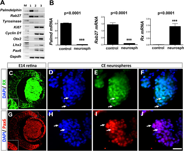
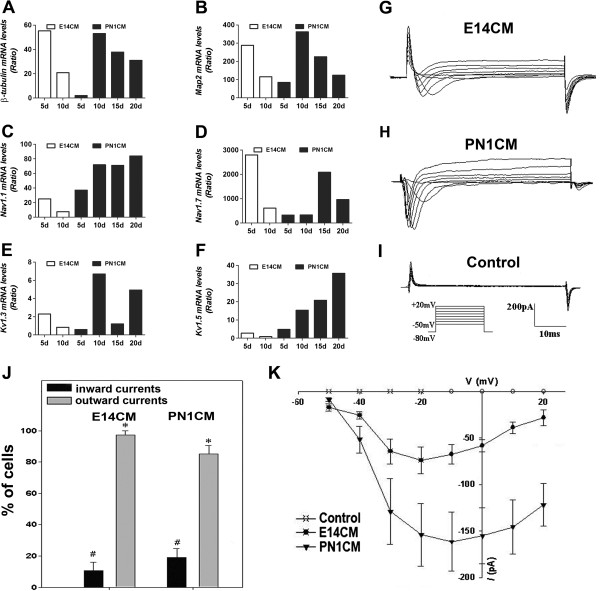
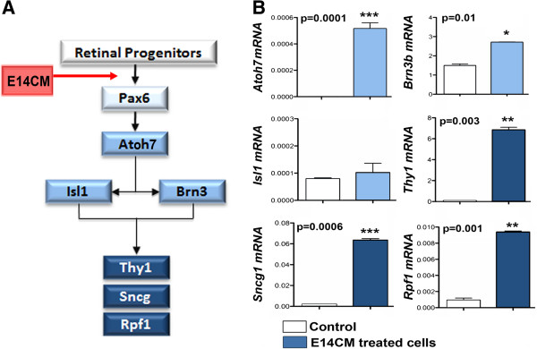
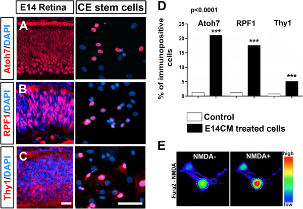
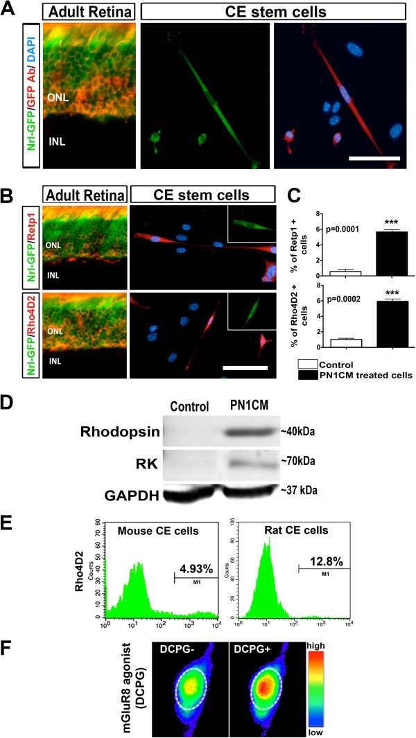
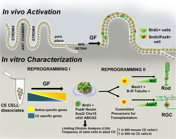
Results: Molecular and cellular analysis was carried out to examine the plasticity of CE stem cells, obtained from mice expressing green fluorescent protein (GFP) under the influence of the promoter of the rod photoreceptor-specific gene, Nrl, using the neurospheres assay. Differentiation was induced by specific culture conditions and evaluated by both transcripts and protein levels of lineage-specific regulators and markers. Temporal pattern of their levels were examined to determine the expression of genes and proteins underlying the regulatory hierarchy of cells specific differentiation in vitro. Functional attributes of differentiation were examined by the presence of current profiles and pharmacological mobilization of intracellular calcium using whole cell recordings and Fura-based calcium imaging, respectively. We demonstrate that stem cells in adult CE not only have the capacity to generate functional neurons, acquiring the expression of sodium and potassium channels, but also respond to specific cues in culture and preferentially differentiate along the lineages of retinal ganglion cells (RGCs) and rod photoreceptors, the early and late born retinal neurons, respectively. The retinal differentiation of CE stem cells was characterized by the temporal acquisition of the expression of the regulators of RGCs and rod photoreceptors, followed by the display of cell type-specific mature markers and mobilization of intracellular calcium.

Conclusions: Our study demonstrates the bonafide retinal potential of adult CE stem cells and suggests that their plasticity could be harnessed for clinical purposes once barriers associated with any lineage conversion, i.e., low efficiency and fidelity is overcome through the identification of conducive culture conditions.


Figures

Figure 1
Figure 1
Cells in CE neurospheres display retinal progenitor properties. Adult CE cells were cultured in the presence of mitogens to generate neurospheres and the acquisition of retinal properties was examined. RT-PCR revealed that the levels of CE-specific transcripts (Tyrosinase, Palmdelphin, and Rab27) decreased in neurospheres with the time (lane 1 = untreated CE cells, lane 2 = 3 days old neurospheres, lane 3 = 6 days old neurospheres). In contrast, cell proliferation-(Ki67 and Cyclin D1,) and retinal progenitor (Otx2, Lhx2 and Pax6)-specific transcript levels increased temporally in neurospheres (A). Q-PCR analysis corroborated the decrease in the levels of Palmdelphin, and Rab27 transcripts and detected Rx transcripts in neurospheres (B). Immunofluorescence analysis revealed that cells in CE neurospheres were immunoreactive for Rx (D, E, F) and Pax6 (H, I, J) as retinal progenitors in E14 retina (C and G). Scale = 50 μm.
Figure 2
Figure 2
Cells in CE neurospheres differentiate into functional neurons. Neurospheres generated by CE stem cells were cultured in the presence of E14CM/PN1CM, and their differentiation into generic neurons was examined. Q-PCR analysis revealed temporal patterns in the acquisition of the expression of neuron-specific marker (β − tubulin, p < 0.0001; and Map2, p < 0.0001) (A, B), tetrodotoxin-sensitive sodium channel (NaV1.1, p = 0.0001; and NaV1.7, p < 0.0001) (C, D), and potassium channels α subunit (Kv1.3, p < 0.0001; and Kv1.5, p = 0.004) (E, F) genes in E14/PN1CM. The levels represent the expression, relative to that in untreated CE cells (ratio). Whole cell voltage clamp recordings revealed fast inward currents in 10.8% (N = 37) of cells in E14CM (G, J) and 19.5% (N = 47) of cells in PN1CM (H, J). The current-voltage (I-V) curve (K) exhibited a typical I-V relationship of voltage gated Na + channels. Cells in both conditions (>80%; N = 37) displayed sustained outward currents conducted most likely by outwardly rectifying K + channels. These currents were not detected in control CE cells (I, K).
Figure 3
Figure 3
Cells in CE neurospheres activate RGC-specific regulatory gene expression. CE neurospheres were cultured in the presence of E14CM for 5 days and RGC regulatory gene expression [37] was examined (A). Q-PCR analysis of cells revealed a significant increase in levels of transcripts corresponding to the regulators of differentiation (Atoh7 and Brn3b) and maturation (Rpf1, Thy1, and Sncg) of RGCs in differentiation conditions, compared to controls (B). However, no significant induction in the expression of Isl1 was observed. Controls: CE untreated cells.
Figure 4
Figure 4
Cells in CE neurospheres express RGC-specific regulatory and marker proteins. Immunofluorescence analysis of CE cells in the presence of E14CM for 5 days revealed a subset expressing immunoreactivities corresponding to RGC markers, Atoh7, RPF1 and Thy1, like RGC progenitors/precursors in E14 retina (A-C), the proportion of which was significantly higher, compared to controls (D). Calcium imaging by Fura2 revealed the mobilization of intracellular calcium in differentiated cells in the presence of NMDA agonist (NMDA+) and not in its absence (NMDA-), indicating the presence of ionotropic glutamate NMDA receptor expressed by RGCs in vivo(E). Bar illustrates fluorescence intensity in a pseudo-color scale (E). Controls: CE untreated cells. Scale = 50 μm.
Figure 5
Figure 5
Cells in CE neurospheres activate rod photoreceptor-specific regulatory gene expression. CE neurospheres were cultured in the presence of PN1CM for 20 days and rod photoreceptor regulatory gene expression [42] was examined (A). Q-PCR analysis revealed a temporal increase in levels of genes corresponding to the regulators of rod differentiation (Crx, Nrl, and Nr2e3) and maturation (Rhodopsin kinase, Rhodopsin, Gnat1, Phosducin, Recoverin, and Arrestin) in differentiation conditions, compared to controls (B). C = control (CE untreated cells).
Figure 6
Figure 6
Cells in CE neurospheres cells express rod photoreceptor-specific regulatory and marker proteins. After CE neurospheres were cultured in the presence of PN1CM for 20 days, a small subset of cells was positive of GFP fluorescence, indicating the activation of Nrl promoter (A). A selected field shows three GFP-positive cells immunoreactive to GFP antibody demonstrating the specificity of the Nrl-GFP-fluorescence, similar to that in the section of PN1 Nrl-GFP mouse retina. That the rare Nrl-GFP-positive cells were of rod photoreceptor lineage was demonstrated by co-localization of rhodopsin immunoreactivities, detected by RetP1 (upper panel) and Rho4D2 (lower panel) with Nrl-GFP fluorescence, as in PN1 Nrl-GFP retinal sections (B). The proportion of cells with rhodopsin immunoreactivities, detected by RetP1 and Rho4D2, was significantly higher in cells in differentiation conditions than in controls (C). Western analysis of cells after 20 days of differentiation revealed 40 kD and 70 kD bands, immunoreactive to Rhodopsin and Rhodopsin Kinase (RK), respectively (D). Examination of species-specific difference in the retinal potential of CE cell revealed mouse and rat CE neurospheres, subjected to identical culture in PN1CM, generate 4.93% and 12.8% of Rho4D2 positive photoreceptors, respectively, on FACS analysis (E). Calcium imaging by Fura2 revealed the mobilization of intracellular calcium by differentiated cells in the presence of agonist DCPG (DCPG+) and not in its absence (DCPG-), demonstrating the presence of mGluR8 metabotropic glutamate receptor, expressed by rod photoreceptors in vivo(F). Controls: CE untreated cells. Bar illustrates fluorescence intensity in a pseudo-color scale. ONL = outer nuclear layer; INL = inner nuclear layer. Scale = 50 μm.
Figure 7
Figure 7
Schematic representation of putative re-programming of CE stem cells in vitro.In vivo exposure of adult CE to exogenous growth factors (GF) such as FGF2 and insulin promotes proliferation of quiescent CE stem cells and some of them express Pax6 [45]. Whether or not these cells are capable of generating neurons or more specifically retinal cells is not well known. These cells, when cultured in the presence of mitogens, generate neurospheres accompanied by a decrease in the expression of CE-specific genes and the acquisition of the expression of genes corresponding to retinal progenitors, representing the first stage of reprogramming. The resulting neurospheres consists of heterogeneous population of cells with subsets, which are BrdU positive, and express pan neural and retinal progenitor markers. The frequency of CE cells capable of generating neurospheres is 1 in 600 mouse and 500 rat CE cells, as determined by LDA analyses [1,4]. Cells in neurospheres have the potential to respond to specific cues for retinal differentiation. Therefore, depending upon the cues, they activate expression of regulatory genes for rod photoreceptors (e.g., Nrl) or RGCs (e.g., Atoh7) to differentiate along specific retinal sub-lineages, representing the second stage of reprogramming. The efficiency and fidelity of retinal lineage conversion will be influenced by the efficiency of reprogramming at both stages. Since the reprogramming is non-cell autonomous, its efficiency will directly depend upon culture conditions, the variability in which might have led to contradictory results.

Similar articles

Cited by

References

    1. Tropepe V, Coles BL, Chiasson BJ, Horsford DJ, Elia AJ, McInnes RR, van der Kooy D. Retinal stem cells in the adult mammalian eye. Science. 2000;287(5460):2032–2036. doi: 10.1126/science.287.5460.2032. - DOI - PubMed
    1. Ahmad I, Tang L, Pham H. Identification of neural progenitors in the adult mammalian eye. Biochem Biophys Res Commun. 2000;270(2):517–521. doi: 10.1006/bbrc.2000.2473. - DOI - PubMed
    1. Das AV, James J, Zhao X, Rahnenführer J, Ahmad I. Identification of c-Kit receptor as a regulator of adult neural stem cells in the mammalian eye: interactions with Notch signaling. Dev Biol. 2004;273(1):87–105. doi: 10.1016/j.ydbio.2004.05.023. - DOI - PubMed
    1. Das AV, James J, Rahnenführer J, Thoreson WB, Bhattacharya S, Zhao X, Ahmad I. Retinal properties and potential of the adult mammalian ciliary epithelium stem cells. Vision Res. 2005;45(13):1653–1666. doi: 10.1016/j.visres.2004.12.017. - DOI - PubMed
    1. Das AV, Zhao X, James J, Kim M, Cowan KH, Ahmad I. Neural stem cells in the adult ciliary epithelium express GFAP and are regulated by Wnt signaling. Biochem Biophys Res Commun. 2006;339(2):708–716. doi: 10.1016/j.bbrc.2005.11.064. - DOI - PubMed

Publication types

LinkOut - more resources