

In vivo motion of femoral condyles during weight-bearing flexion after anterior cruciate ligament rupture using biplane radiography
- PMID: 24149168
- PMCID: PMC3772605
In vivo motion of femoral condyles during weight-bearing flexion after anterior cruciate ligament rupture using biplane radiography
Abstract
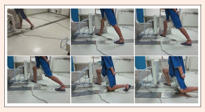
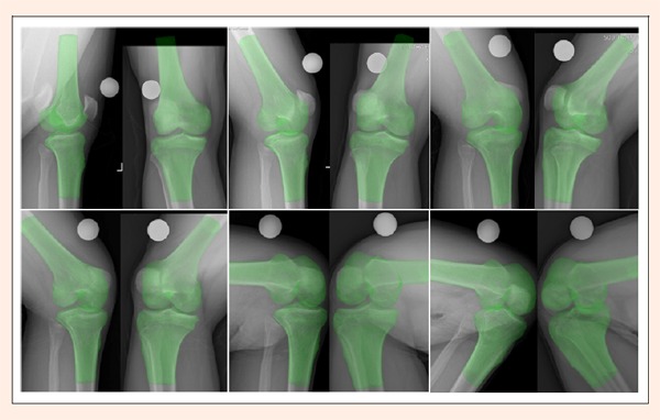
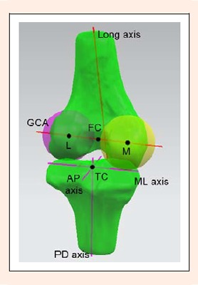
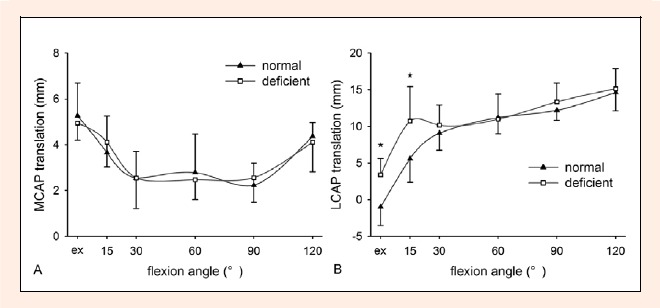
The purpose of this study was to investigate in vivo three- dimensional tibiofemoral kinematics and femoral condylar motion in knees with anterior cruciate ligament (ACL) deficiency during a knee bend activity. Ten patients with unilateral ACL rupture were enrolled. Both the injured and contralateral normal knees were imaged using biplane radiography at extension and at 15°, 30°, 60°, 90°, and 120° of flexion. Bilateral knees were next scanned by computed tomography, from which bilateral three-dimensional knee models were created. The in vivo tibiofemoral motion at each flexion position was reproduced through image registration using the knee models and biplane radiographs. A joint coordinate system containing the geometric center axis of the femur was used to measure the tibiofemoral motion. In ACL deficiency, the lateral femoral condyle was located significantly more posteriorly at extension and at 15° (p < 0.05), whereas the medial condylar position was changed only slightly. This constituted greater posterior translation and external rotation of the femur relative to the tibia at extension and at 15° (p < 0.05). Furthermore, ACL deficiency led to a significantly reduced extent of posterior movement of the lateral condyle during flexion from 15° to 60° (p < 0.05). Coupled with an insignificant change in the motion of the medial condyle, the femur moved less posteriorly with reduced extent of external rotation during flexion from 15° to 60° in ACL deficiency (p < 0.05). The medial- lateral and proximal-distal translations of the medial and lateral condyles and the femoral adduction-abduction rotation were insignificantly changed after ACL deficiency. The results demonstrated that ACL deficiency primarily changed the anterior-posterior motion of the lateral condyle, producing not only posterior subluxation at low flexion positions but also reduced extent of posterior movement during flexion from 15° to 60°. Key PointsThree-dimensional tibiofemoral kinematics and femoral condylar motion in ACL-deficient knees during upright weight-bearing flexion were measured using biplane radiography with the geometric center axis.ACL deficiency caused posterior subluxation of the lateral condyle with excess external femoral rotation at early flexion positions.On flexion from 15° to 60°, the lateral condyle moved slightly posteriorly in ACL deficiency leading to reduced extent of external femoral rotation.
Keywords: anterior cruciate ligament; femoral condyle; injury; kinematics; radiography; tibiofemoral.
Figures
Similar articles
-
[Anterior Cruciate Ligament Tears - Influence on Terminal Extension].Acta Chir Orthop Traumatol Cech. 2018;85(1):22-28. Acta Chir Orthop Traumatol Cech. 2018. PMID: 30257765 Czech.
-
In vivo kinematics and ligamentous function of the knee during weight-bearing flexion: an investigation on mid-range flexion of the knee.Knee Surg Sports Traumatol Arthrosc. 2020 Mar;28(3):797-805. doi: 10.1007/s00167-019-05499-y. Epub 2019 Apr 10. Knee Surg Sports Traumatol Arthrosc. 2020. PMID: 30972464 Free PMC article.
-
MRI analysis of in vivo meniscal and tibiofemoral kinematics in ACL-deficient and normal knees.J Orthop Res. 2006 Jun;24(6):1208-17. doi: 10.1002/jor.20139. J Orthop Res. 2006. PMID: 16652339
-
Three-dimensional in vivo dynamic motion analysis of anterior cruciate ligament-deficient knees during squatting using geometric center axis of the femur.J Orthop Sci. 2016 Mar;21(2):159-65. doi: 10.1016/j.jos.2015.11.001. Epub 2015 Dec 20. J Orthop Sci. 2016. PMID: 26714666
-
In vivo kinematics of medial unicompartmental osteoarthritic knees during activities of daily living.Knee. 2014;21 Suppl 1:S10-4. doi: 10.1016/S0968-0160(14)50003-8. Knee. 2014. PMID: 25382361 Review.
Cited by
-
The Relationship between Anterior Cruciate Ligament Injury and Osteoarthritis of the Knee.Adv Orthop. 2015;2015:928301. doi: 10.1155/2015/928301. Epub 2015 Apr 19. Adv Orthop. 2015. PMID: 25954533 Free PMC article. Review.
-
In vivo posterior cruciate ligament elongation in running activity after anatomic and non-anatomic anterior cruciate ligament reconstruction.Knee Surg Sports Traumatol Arthrosc. 2017 Apr;25(4):1177-1183. doi: 10.1007/s00167-016-4180-4. Epub 2016 Jun 2. Knee Surg Sports Traumatol Arthrosc. 2017. PMID: 27256277 Free PMC article.
-
Identifying the Functional Flexion-extension Axis of the Knee: An In-Vivo Kinematics Study.PLoS One. 2015 Jun 3;10(6):e0128877. doi: 10.1371/journal.pone.0128877. eCollection 2015. PLoS One. 2015. PMID: 26039711 Free PMC article.
-
Influence of leg axis alignment on MRI T2* mapping of the knee in young professional soccer players.BMC Musculoskelet Disord. 2024 Feb 15;25(1):144. doi: 10.1186/s12891-024-07233-3. BMC Musculoskelet Disord. 2024. PMID: 38360606 Free PMC article.
-
Double-bundle anterior cruciate ligament reconstruction improves tibial rotational instability: analysis of squatting motion using a 2D/3D registration technique.J Orthop Surg Res. 2018 May 16;13(1):111. doi: 10.1186/s13018-018-0825-y. J Orthop Surg Res. 2018. PMID: 29769139 Free PMC article.
References
-
- Ahn J.H., Bae T.S., Kang K.S., Kang S.Y., Lee S.H. (2011) Longitudinal tear of the medial meniscus posterior horn in the anterior cruciate ligament-deficient knee significantly influences anterior stability. American Journal of Sports Medicine 39(10), 2187-2193 - PubMed
-
- Allen C.R., Wong E.K., Livesay G.A., Sakane M., Fu F.H., Woo S.L. (2000) Importance of the medial meniscus in the anterior cruciate ligament-deficient knee. Journal of Orthopaedic Research 18(1), 109-115 - PubMed
-
- Asano T., Akagi M., Tanaka K., Tamura J., Nakamura T. (2001) In vivo three-dimensional knee kinematics using a biplanar image-matching technique. Clinical Orthopaedics and Related Research 388, 157-166 - PubMed
-
- Brandsson S., Karlsson J., Eriksson B.I., Karrholm J. (2001) Kinematics after tear in the anterior cruciate ligament: dynamic bilateral radiostereometric studies in 11 patients. Acta Orthopaedica Scandinavica 72(4), 372-378 - PubMed
-
- Defrate L.E., Papannagari R., Gill T.J., Moses J.M., Pathare N.P., Li G. (2006) The 6 degrees of freedom kinematics of the knee after anterior cruciate ligament deficiency: an in vivo imaging analysis. American Journal of Sports Medicine 34(8), 1240-1246 - PubMed
LinkOut - more resources
Full Text Sources