. 2013 Dec;8(12):1373-83.
doi: 10.4161/epi.26781. Epub 2013 Oct 22.

Epigenetic regulation of DACH1, a novel Wnt signaling component in colorectal cancer

Affiliations

Epigenetic regulation of DACH1, a novel Wnt signaling component in colorectal cancer

Wenji Yan et al. Epigenetics. 2013 Dec.

Abstract

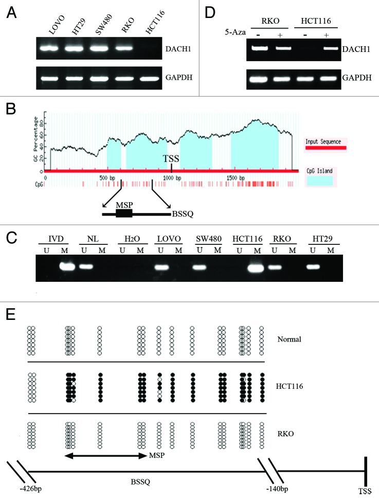
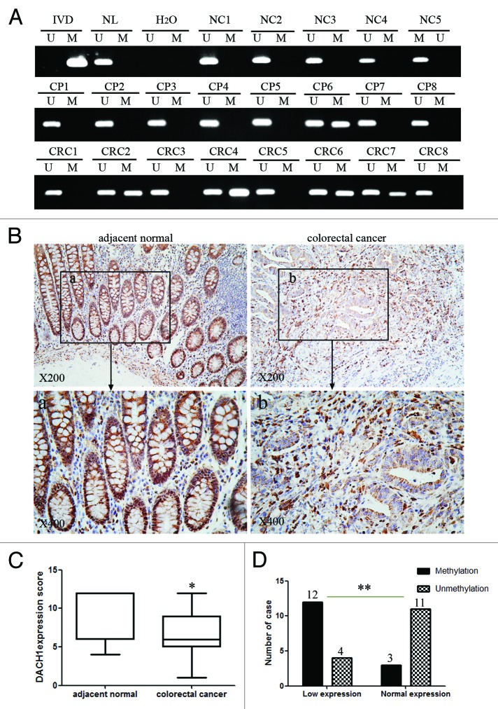
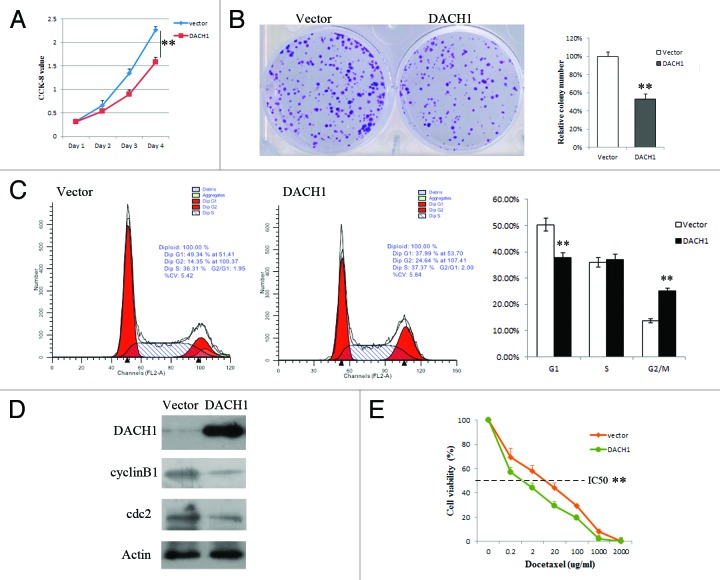
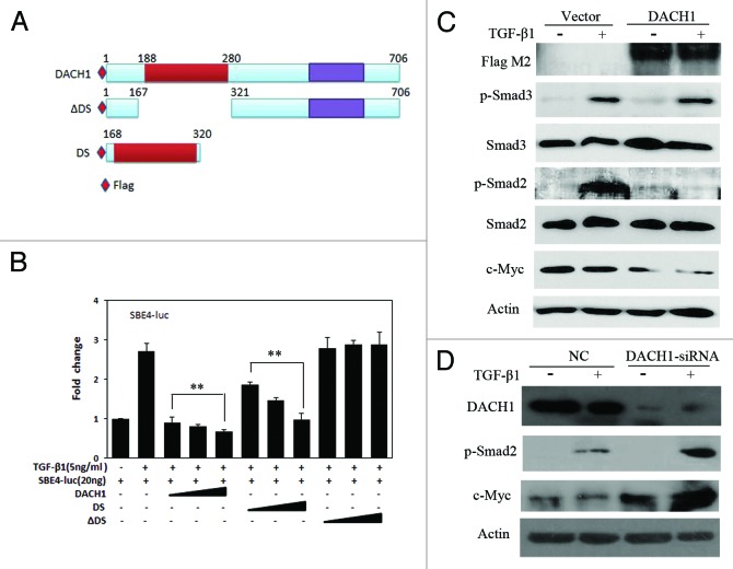
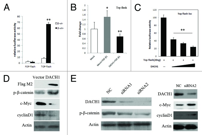
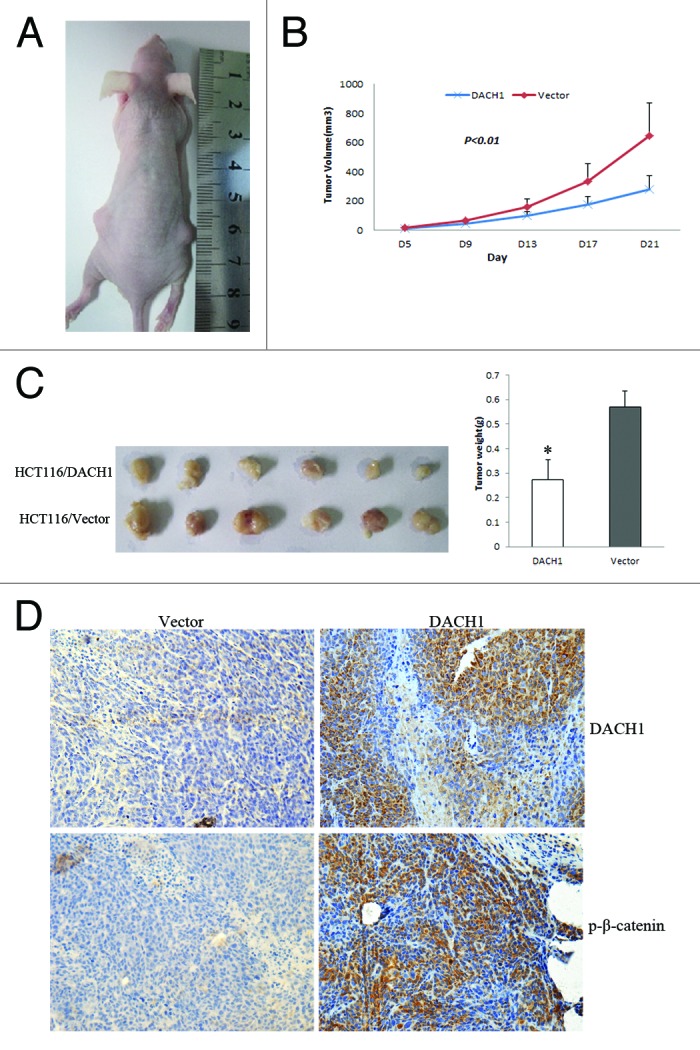
Colorectal cancer (CRC) is one of the common malignant tumors worldwide. Both genetic and epigenetic changes are regarded as important factors of colorectal carcinogenesis. Loss of DACH1 expression was found in breast, prostate, and endometrial cancer. To analyze the regulation and function of DACH1 in CRC, 5 colorectal cancer cell lines, 8 cases of normal mucosa, 15 cases of polyps and 100 cases of primary CRC were employed in this study. In CRC cell lines, loss of DACH1 expression was correlated with promoter region hypermethylation, and re-expression of DACH1 was induced by 5-Aza-2'-deoxyazacytidine treatment. We found that DACH1 was frequently methylated in primary CRC and this methylation was associated with reduction in DACH1 expression. These results suggest that DACH1 expression is regulated by promoter region hypermethylation in CRC. DACH1 methylation was associated with late tumor stage, poor differentiation, and lymph node metastasis. Re-expression of DACH1 reduced TCF/LEF luciferase reporter activity and inhibited the expression of Wnt signaling downstream targets (c-Myc and cyclinD1). In xenografts of HCT116 cells in which DACH1 was re-expressed, tumor size was smaller than in controls. In addition, restoration of DACH1 expression induced G2/M phase arrest and sensitized HCT116 cells to docetaxel. DACH1 suppresses CRC growth by inhibiting Wnt signaling both in vitro and in vivo. Silencing of DACH1 expression caused resistance of CRC cells to docetaxel. In conclusion, DACH1 is frequently methylated in human CRC and methylation of DACH1 may serve as detective and prognostic marker in CRC.

Keywords: DACH1; DNA methylation; Wnt signaling; chemosensitivity; colorectal cancer.


Figures

None
Figure 1. Expression of DACH1 is downregulated by DNA methylation in colorectal cancer cell lines. (A) Expression of DACH1 was analyzed by semi-quantitative RT-PCR in colorectal cancer cell lines (LOVO, HT29, SW480, RKO, and HCT116). (B) CpG islands of the DACH1 gene locus predicted by “MethPrimer” (http://www.urogene.org/cgi-bin/methprimer/methprimer.cgi). CpG sites between -1000 and +1000 bp from the transcription start site (TSS) are presented by vertical bars. Sequence of methylation specific PCR (MSP) and bisulfite sequencing (BSSQ) products is shown. (C) Methylation status of the DACH1 promoter region in colorectal cancer cell lines. Primer efficiency was verified by positive control (in vitro methylated DNA, IVD) and negative control (normal blood lymphocyte DNA, NL). U indicates the presence of unmethylated alleles; M indicates the presence of methylated alleles. (D) Expression of DACH1 was analyzed by semi-quantitative RT-PCR in HCT116 and RKO cell lines in the absence or presence of 2 μmol/L 5-Aza (+) for 96 h. (E) Bisulfite sequencing of DACH1 was performed in HCT116, RKO cell lines, and normal colon mucosa. The region of CpG island studied by MSP is indicated by a double-headed arrow, spanning 130 bp. Filled circles represent methylated CpG sites within the DACH1 CpG island, and open circles denote unmethylated CpG sites. Bisulfite sequencing focused on a 286 bp (-426 bp to -140 bp) CpG island upstream of the DACH1 TST.
None
Figure 2. DNA methylation and expression of DACH1 in primary colorectal cancer. (A) Representative MSP results of DACH1 methylation status in normal colon mucosa (NC), colorectal adenomatous polyps (CP) and colorectal cancer tissues (CRC). (B) Representative images of DACH1 protein expression in colorectal cancer and their adjacent non-tumor tissues determined by IHC. (upper images, X200; lower images, X400). (C) DACH1 expression scores are shown as box plots, horizontal lines represent the median score; the bottom and top of the boxes representing the 25th and 75th percentiles, respectively; vertical bars represent the range of data. Expression of DACH1 was different between adjacent normal and tumor tissues in 30-matched primary CRCs. *P < 0.05. (D) The correlation between DACH1 hypermethylation and expression level was analyzed in 30-matched primary CRCs (**P < 0.01).
None
Figure 3. Effect of DACH1 expression on tumor growth and cell cycle in HCT116 cells. (A) Growth curves, which represents the effect of DACH1 expression on cell viability in HCT116 cells was tested by CCK-8 kit daily for 96 h. (B) Colony formation assay shows the effect of DACH1 expression on cancer cell growth. The left panel shows the representative images of PCMV6-AC-DACH1-GFP or empty vector (PCMV6-AC-GFP) group. Right panel shows colony numbers (**P < 0.01). (C) Representative cell cycle and flow cytometry data (**P < 0.01). (D) western blot results in HCT116 cells with unexpressed DACH1 and re-expressed DACH1 . (E) The cell viability assay showed the effect of DACH1 restoration on the sensitivity of HCT116 cells to docetaxel (**P < 0.01).
None
Figure 4. Effect of DACH1 on TGF-β signaling pathway. (A) Schematic structure of proteins expressed by DACH1, DS, and ΔDS vector. (B) Luciferase activity was detected in HCT116 cells transfected with empty vector and increasing doses of DACH1, DS, and ΔDS (50 ng, 100 ng, 200 ng/well for DACH1 and the same molecular for DS and ΔDS), with the addition of 5 ng/ml TGF-β1 in each group (**P < 0.01). (C) HCT116 cells were transfected with DACH1 with or without TGF-β1 addition, and indicated proteins were detected with the indicated antibodies by western blot. (D) western blot shows the expression of TGF-β signaling downstream genes before and after DACH1 was knocked down in HT29 cells.
None
Figure 5. DACH1 represses Wnt/β-catenin signaling activity in colorectal cancer cells. (A) β-catenin expression vector was co-transfected with TCF/LEF Topflash reporter or its mutant, Fopflash, into HCT116 cells. Luciferase activity was normalized to Renilla luciferase activity (**P < 0.01). (B) Luciferase activity in mock, mock + TGF-β1, and DACH1 + TGF-β1 groups. (*P < 0.05, **P < 0.01). (C) Effect of increasing dose of DACH1 (50 ng, 100 ng, 200 ng/well) on Wnt signaling activity (**P < 0.01). (D) western blot showing the levels of p-β-catenin and Wnt signaling downstream genes, cyclinD1, and c-Myc, before and after DACH1 transfection in HCT116 cells. (E) western blot of HT29 cells before and after DACH1 was knocked down. The left panel shows the level of p-β-catenin was decreased after knocking down DACH1 in HT29 cells; the right panel shows the expression of c-Myc and cyclinD1, which was increased after knocking down DACH1 in HT29 cells.
None
Figure 6. DACH1 retarded tumor growth in vivo. (A) Representative picture of tumor growth in nude mice subcutaneously inoculated with DACH1 expressing vector (right) and empty vector (left). (B) Subcutaneous tumor growth curve in xenograft mice with or without DACH1 expression. Retarded tumor growth was found in DACH1-expressing group. (C) Histogram represents average weight of xenograft in DACH1-expressing and DACH1-non-expressing groups. (D) Increased p-β-catenin was found in DACH1-expressing xenografts by IHC staining (x400).

Similar articles

Cited by

References

    1. Jemal A, Siegel R, Ward E, Hao Y, Xu J, Thun MJ. Cancer statistics, 2009. CA Cancer J Clin. 2009;59:225–49. doi: 10.3322/caac.20006. - DOI - PubMed
    1. Ferlay J, Shin HR, Bray F, Forman D, Mathers C, Parkin DM. Estimates of worldwide burden of cancer in 2008: GLOBOCAN 2008. Int J Cancer. 2010;127:2893–917. doi: 10.1002/ijc.25516. - DOI - PubMed
    1. Zhang W, Glöckner SC, Guo M, Machida EO, Wang DH, Easwaran H, Van Neste L, Herman JG, Schuebel KE, Watkins DN, et al. Epigenetic inactivation of the canonical Wnt antagonist SRY-box containing gene 17 in colorectal cancer. Cancer Res. 2008;68:2764–72. doi: 10.1158/0008-5472.CAN-07-6349. - DOI - PMC - PubMed
    1. Riojas MA, Guo M, Glöckner SC, Machida EO, Baylin SB, Ahuja N. Methylation-induced silencing of ASC/TMS1, a pro-apoptotic gene, is a late-stage event in colorectal cancer. Cancer Biol Ther. 2007;6:1710–6. doi: 10.4161/cbt.6.11.4829. - DOI - PubMed
    1. Hanada N, Takahata T, Zhou Q, Ye X, Sun R, Itoh J, Ishiguro A, Kijima H, Mimura J, Itoh K, et al. Methylation of the KEAP1 gene promoter region in human colorectal cancer. BMC Cancer. 2012;12:66. doi: 10.1186/1471-2407-12-66. - DOI - PMC - PubMed

Publication types

MeSH terms

LinkOut - more resources