. 2013 Oct 22;4(5):e00801-13.
doi: 10.1128/mBio.00801-13.

Infectious bronchitis virus generates spherules from zippered endoplasmic reticulum membranes

Affiliations

Infectious bronchitis virus generates spherules from zippered endoplasmic reticulum membranes

Helena J Maier et al. mBio. .

Abstract

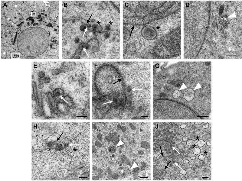
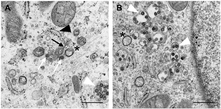
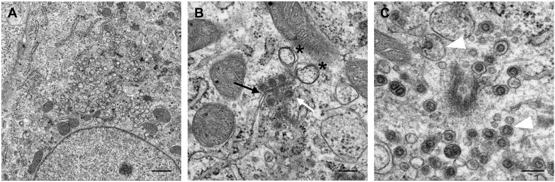
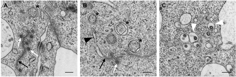
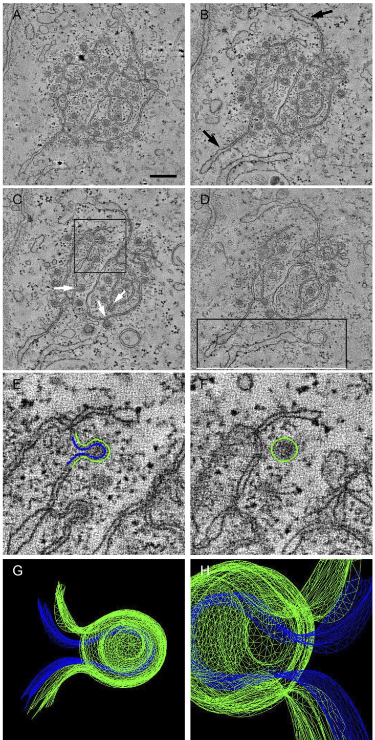
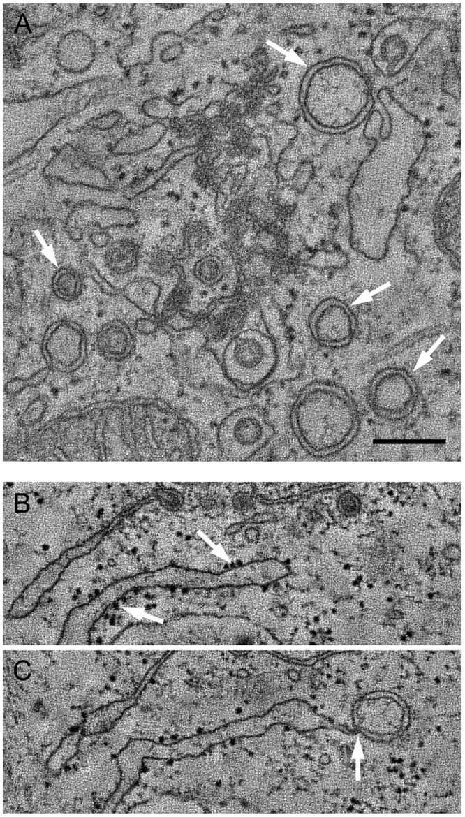
Replication of positive-sense RNA viruses is associated with the rearrangement of cellular membranes. Previous work on the infection of tissue culture cell lines with the betacoronaviruses mouse hepatitis virus and severe acute respiratory syndrome coronavirus (SARS-CoV) showed that they generate double-membrane vesicles (DMVs) and convoluted membranes as part of a reticular membrane network. Here we describe a detailed study of the membrane rearrangements induced by the avian gammacoronavirus infectious bronchitis virus (IBV) in a mammalian cell line but also in primary avian cells and in epithelial cells of ex vivo tracheal organ cultures. In all cell types, structures novel to IBV infection were identified that we have termed zippered endoplasmic reticulum (ER) and spherules. Zippered ER lacked luminal space, suggesting zippering of ER cisternae, while spherules appeared as uniform invaginations of zippered ER. Electron tomography showed that IBV-induced spherules are tethered to the zippered ER and that there is a channel connecting the interior of the spherule with the cytoplasm, a feature thought to be necessary for sites of RNA synthesis but not seen previously for membrane rearrangements induced by coronaviruses. We also identified DMVs in IBV-infected cells that were observed as single individual DMVs or were connected to the ER via their outer membrane but not to the zippered ER. Interestingly, IBV-induced spherules strongly resemble confirmed sites of RNA synthesis for alphaviruses, nodaviruses, and bromoviruses, which may indicate similar strategies of IBV and these diverse viruses for the assembly of RNA replication complexes.

Importance: All positive-sense single-stranded RNA viruses induce rearranged cellular membranes, providing a platform for viral replication complex assembly and protecting viral RNA from cellular defenses. We have studied the membrane rearrangements induced by an important poultry pathogen, the gammacoronavirus infectious bronchitis virus (IBV). Previous work studying closely related betacoronaviruses identified double-membrane vesicles (DMVs) and convoluted membranes (CMs) derived from the endoplasmic reticulum (ER) in infected cells. However, the role of DMVs and CMs in viral RNA synthesis remains unclear because these sealed vesicles lack a means of delivering viral RNA to the cytoplasm. Here, we characterized structures novel to IBV infection: zippered ER and small vesicles tethered to the zippered ER termed spherules. Significantly, spherules contain a channel connecting their interior to the cytoplasm and strongly resemble confirmed sites of RNA synthesis for other positive-sense RNA viruses, making them ideal candidates for the site of IBV RNA synthesis.


Figures

FIG 1
FIG 1
Characterization of virus replication. (A) CK cells were infected with IBV, and total RNA was extracted at hourly intervals. Levels of genomic RNA (gRNA; Ai) and N sgRNA (Aii) were measured by 2-step qRT-PCR. Data from three independent experiments are shown. (B) CK cells were infected with recombinant IBV BeauR-hrLuc-Δ5a expressing Renilla luciferase in place of gene 5a. Protein lysates were harvested at hourly intervals, and Renilla luciferase activity was measured. Data representing the means and standard deviations of the results of three independent experiments are shown. (C) CK cells were infected with IBV and cell supernatants harvested at hourly intervals. Release of progeny virus was determined by a plaque assay. Data representing the means and standard deviations of the results of three independent experiments are shown.
FIG 2
FIG 2
Minor populations of dsRNA and Nsp12 colocalize. (A, B, and C) CK cells were infected with IBV and fixed using 4% paraformaldehyde at 4 (A), 6 (B), or 8 (C) hpi. Cells were labeled with anti-dsRNA (red) and anti-nsp12 (green). Nuclei were stained with DAPI (blue). Bars indicate 10 µm. (D and E) Colocalization of dsRNA (D) and nsp12 (E) was quantified using the Imaris Coloc module. Means and standard deviations of the results of three independent experiments are shown. Ns, not significant; **, P ≤ 0.01 (Student’s t test).
FIG 3
FIG 3
Zippered ER, spherules, and double-membrane vesicles are induced by IBV in CK cells. CK cells were infected with IBV, and at 16 hpi (A to I) or 24 hpi (J), cells were fixed by high-pressure freezing (A to G) or chemical fixation (H and J). Double-membrane vesicles are marked with asterisks, spherules are marked with white arrows, zippered ER is marked with black arrows, and virus particles in vesicles are marked with white arrowheads. The white boxes in panel A highlight regions of IBV-induced membrane rearrangements. The region marked by the lower box is expanded in panel B. Scale bars: A, 2 µm; B and J, 200 nm; C, E, and F, 100 nm; D and G to I, 500 nm.
FIG 4
FIG 4
Zippered ER, spherules, and double-membrane vesicles are induced by IBV in Vero cells. Vero cells were infected with IBV, and at 16 hpi, cells were chemically fixed. Double-membrane vesicles are marked with asterisks, spherules are marked with white arrows, zippered ER is marked with black arrows, virus particles in vesicles are marked with white arrowheads, and ribosomes are marked with black arrowheads. Scale bars indicate 100 nm (A) or 200 nm (B).
FIG 5
FIG 5
IBV-induced membrane rearrangements can be detected at 7 hpi. CK cells were infected with IBV and chemically fixed at hourly intervals. Double-membrane vesicles (*), spherules (white arrow), and zippered ER (black arrow), as well as virus particles in vesicles (white arrowhead), were detectable at 7 hpi. Scale bars indicate 1 µm (A) or 200 nm (B and C).
FIG 6
FIG 6
IBV infection induced zippered ER, spherules, and double-membrane vesicles in ex vivo tracheal organ culture (TOC). TOCs were infected with IBV and at 24 hpi were chemically fixed. Double-membrane vesicles (*), spherules (white arrow), and zippered ER (black arrow) (A and B), as well as virus particles in vesicles (white arrowhead) (C), could be detected. Ribosomes could be detected on ER membranes (B, black arrowhead). Scale bars indicate 200 nm.
FIG 7
FIG 7
Spherules are derived from and connected to the zippered ER and have a channel connecting their interior to the cytoplasm. CK cells infected with IBV were chemically fixed at 16 hpi, and a dual-axis electron tomography data set was collected from a 300-nm-thick section. Images were collected at 1° intervals over a ±60° range using FEI T20 200 kV TEM and Acquire 3D software. Reconstruction and modeling were carried out using IMOD software. (A to D) Four images taken from the tomogram (for the full tomogram, see Movie S1 in the supplemental material). Collapsed ER membranes (arrows) (B) fold to form individual spherules with a clear channel connecting the cytoplasm and spherule interior (arrows indicate spherule necks) (C) and a connection between an ER tubule and a DMV (D). The area highlighted in panel C was modeled by tracing the two membranes in consecutive slices (outer membrane in green, inner membrane in blue). (E to F) Two images from the contour data set (for the full contour data set, see Movie S2). The three-dimensional mesh model of this spherule, represented by two images (G to H), clearly displays the open channel between the cell cytoplasm and the spherule center. For the model movie, see Movie S3. The scale bar in panel A indicates 50 nm.
FIG 8
FIG 8
Double-membrane vesicles exist as single vesicles. (A) The tomographic reconstruction of an IBV-infected cell chemically fixed at 16 hpi shows an accumulation of individual DMVs which do not appear to be connected (arrows). For the full reconstruction, see Movie S4 in the supplemental material. The data set represented by panel A was collected under the same conditions as described for Fig. 7. (B and C) Double-membrane vesicles appear to be connected to rough ER. Ribosomes on the ER tubule are highlighted in panel B (arrows), and the connection between the ER tubule and DMV is demonstrated in panel C (arrow). Panels B and C represent still images taken from Movie S6, which in turn was extracted from the full tomogram represented by Movie S1, focusing on the area highlighted in Fig. 7D. The scale bar in panel A indicates 50 nm.

Comment in

References

    1. Miller S, Krijnse-Locker J. 2008. Modification of intracellular membrane structures for virus replication. Nat. Rev. Microbiol. 6:363–374 - PMC - PubMed
    1. Denison MR. 2008. Seeking membranes: positive-strand RNA virus replication complexes. PLoS Biol. 6:e270.10.1371/journal.pbio.0060270 - DOI - PMC - PubMed
    1. Paul D, Bartenschlager R. 2013. Architecture and biogenesis of plus-strand RNA virus replication factories. World J. Virol. 2:32–48 - PMC - PubMed
    1. Cottam E, Pierini R, Roberts R, Wileman T. 2009. Origins of membrane vesicles generated during replication of positive-strand RNA viruses. Future Virol. 4:473–485
    1. Gosert R, Egger D, Lohmann V, Bartenschlager R, Blum HE, Bienz K, Moradpour D. 2003. Identification of the hepatitis C virus RNA replication complex in huh-7 cells harboring subgenomic replicons. J. Virol. 77:5487–5492 - PMC - PubMed

Publication types

MeSH terms

LinkOut - more resources