. 2014 Jan;10(1):32-44.
doi: 10.4161/auto.26508. Epub 2013 Jan 1.

Mesenchymal stem cells enhance autophagy and increase β-amyloid clearance in Alzheimer disease models

Affiliations

Mesenchymal stem cells enhance autophagy and increase β-amyloid clearance in Alzheimer disease models

Jin Young Shin et al. Autophagy. 2014 Jan.

Abstract

Current evidence suggests a central role for autophagy in Alzheimer disease (AD), and dysfunction in the autophagic system may lead to amyloid-β (Aβ) accumulation. Using in vitro and in vivo AD models, the present study investigated whether mesenchymal stem cells (MSCs) could enhance autophagy and thus exert a neuroprotective effect through modulation of Aβ clearance In Aβ-treated neuronal cells, MSCs increased cellular viability and enhanced LC3-II expression compared with cells treated with Aβ only. Immunofluorescence revealed that MSC coculture in Aβ-treated neuronal cells increased the number of LC3-II-positive autophagosomes that were colocalized with a lysosomal marker. Ultrastructural analysis revealed that most autophagic vacuoles (AVs) in Aβ-treated cells were not fused with lysosomes, whereas a large portion of autophagosomes were conjoined with lysosomes in MSCs cocultured with Aβ-treated neuronal cells. Furthermore, MSC coculture markedly increased Aβ immunoreactivity colocalized within lysosomes and decreased intracellular Aβ levels compared with Aβ-treated cells. In Aβ-treated animals, MSC administration significantly increased autophagosome induction, final maturation of late AVs, and fusion with lysosomes. Moreover, MSC administration significantly reduced the level of Aβ in the hippocampus, which was elevated in Aβ-treated mice, concomitant with increased survival of hippocampal neurons. Finally, MSC coculture upregulated BECN1/Beclin 1 expression in AD models. These results suggest that MSCs significantly enhance autolysosome formation and clearance of Aβ in AD models, which may lead to increased neuronal survival against Aβ toxicity. Modulation of the autophagy pathway to repair the damaged AD brain using MSCs would have a significant impact on future strategies for AD treatment.

Keywords: Alzheimer disease; BECN1; amyloid beta; autophagy; mesenchymal stem cell.


Figures
None
Figure 1. Aβ induces autophagosome formation in SH-SY5Y cells. SH-SY5Y cells were exposed to various concentration of Aβ (Aβ1–42) for 24 or 72 h. Cell viability was decreased in a dose- and time-dependent manner (A and B). However, reverse peptide Aβ42–1 treatment (20 μM for 24 and 72 h) did not lead to morphological changes or loss of cell viability. Aβ treatment enhanced the autophagy marker (LC3). Western blot analysis showed that Aβ treatment increased expression of LC3-II in a dose- and time-dependent manner (C and D). Values are expressed as mean ± SDs, n = 3. *P < 0.05 for Aβ-treated SH-SY5Y cells vs. untreated SH-SY5Y cells; # P < 0.05 for Aβ-treated SH-SY5Y cells at 24 h vs. Aβ-treated SH-SY5Y cells at 72 h.
None
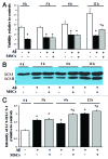
Figure 2. MSCs increase cellular viability and enhance autophagy induction in Aβ-treated SH-SY5Y cells. Aβ-treated SH-SY5Y cells were cocultured with MSCs for 3, 6, 12, or 24 h. MSC coculture significantly increased the viability of Aβ-treated SH-SY5Y cells at 12 h compared with baseline (A). Additionally, MSCs enhanced LC3 expression in Aβ-treated cells compared with Aβ-treated cells. Bar charts illustrate quantification of LC3-II/ LC3-I (B and C). Values are expressed as mean ± SDs, n = 3. *P < 0.05 for Aβ-treated SH-SY5Y cells vs. untreated SH-SY5Y cells; # P < 0.05 for Aβ-treated SH-SY5Y cells vs. MSCs-cocultured SH-SY5Y cells.
None
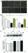
Figure 3. MSCs enhance autolysosome formation. Immunofluorescence showed that MSCs cocultured in Aβ-treated SH-SY5Y cells increased the number of LC3-positive autophagosomes (FITC; green) that were colocalized with lysosomes (Cy3; red) at 6 and 12 h compared with Aβ-treated SH-SY5Y cells (A). A quantitative analysis of FITC and Cy3 double-positive puncta per cell in Aβ-treated SH-SY5Y cells showed that MSC coculture significantly enhanced autolysosome formation compared with Aβ-treated cells (B), data are the mean values of 3 independent experiments, each with no fewer than 100 cells). Additionally, MSC coculture increased lysosomal activity in Aβ-treated SH-SY5Y cells (C), and CQ treatment significantly attenuated cell survival, which was increased when cocultured with MSCs (D). Values are expressed as mean ± SDs, n = 3. *P < 0.05 for Aβ-treated SH-SY5Y cells vs. untreated SH-SY5Y cells; # P < 0.05 for Aβ-treated SH-SY5Y cells vs. MSC-cocultured SH-SY5Y cells; § P < 0.05 for MSC-cocultured SH-SY5Y cells vs. CQ and MSC-cocultured SH-SY5Y cells. Scale bars: 5 μm.
None
Figure 4. MSCs enhance autophagy modulation in CHO cells, stably expressing human APP695. CHO cells were cocultured with MSCs for 6 h. Coculture with MSCs significantly increased expression of LC3 and RAB7 in CHO cells compared with cells without MSCs (A and C). Bar charts illustrate quantification of the LC3-I/II ratio and relative RAB7 expression (B and D). Additionally, coculturing CHO cells with MSCs tended to decrease in intracellular Aβ levels, however, the difference did not reach statistical significance (E). Values are expressed as mean ± SDs (SD), n = 3. *P < 0.05 for CHO cells vs. MSCs cocultured CHO cells.
None
Figure 5. Ultrastructural analyses reveal that MSCs upregulate autolysosome formation in Aβ-treated SH-SY5Y cells. Autophagosomes were rarely noted in SH-SY5Y cells without Aβ treatment (A). Most autophagic vesicles observed in Aβ-treated SH-SY5Y cells did not fuse with lysosomes (B and C); however, MSC coculture led to a large portion of autophagosomes being conjoined with lysosomes (D and E). Yellow arrows indicate autophagosomes and red arrows indicate autolysosomes. Scale bars: 10,000 nm.
None
Figure 6. MSCs enhance Aβ clearance through the autophagy-lysosomal pathway. We assessed Aβ colocalized in lysosomes and its intracellular concentrations to determine whether MSCs-induced autophagy enhanced Aβ clearance. Immunofluorescence analysis of Aβ (green) and LC3 (red) double-positive puncta per cell showed that Aβ within autophagosomes was not changed in SH-SY5Y cells cocultured with MSCs compared with Aβ-treated cells (A and B). However, MSCs cocultured in Aβ-treated cells increased Aβ immunoreactivity (green) that was colocalized in lysosomes (red) compared with Aβ-treated cells (C and D). MSC coculture significantly decreased intracellular Aβ levels in Aβ-treated SH-SY5Y cells, where Aβ concentration was gradually increased in a time-dependent manner (E). Data are the mean values of 3 independent experiments, each with no fewer than 100 cells. Values are expressed as mean ± SDs, n = 3. *P < 0.05 for Aβ-treated SH-SY5Y cells vs. untreated SH-SY5Y cells; # P < 0.05 for Aβ-treated SH-SY5Y cells vs. MSCs-cocultured SH-SY5Y cells. Scale bars: 5 μm.
None
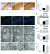
Figure 7. MSCs exert neuroprotective effects in an AD animal model by enhancing degradation of Aβ through autophagy. RBFOX3-positive cells and Nissl-stained cells in the hippocampus were markedly decreased in Aβ-treated mice compared with the controls (A and B), whereas MSC administration markedly increased the survival of RBFOX3-positive and Nissl-stained cells in the brain (C). On stereological analysis, RBFOX3-positive cells in the hippocampus were significantly decreased in in Aβ-treated mice compared with the controls (A and B), whereas MSC administration markedly increased the survival of hippocampal neurons in the AD animal brain (D). After MSC administration in AD animals significantly increased the ratio of LC3-II/LC3-I (E) and RAB7 expression (F). Additionally, MSC administration reduced Aβ immunoreactivity (G to I) and the level of Aβ level (J) compared with only-Aβ-treated mice. Ultrastructural analyses showed that autophagosomes were rarely noted in normal mice (K to M), whereas autophagic vesicles that were not fused with lysosomes were abnormally accumulated in Aβ-treated mice (N to P). In contrast, MSC administration induced a large portion of autophagosomes that were fused with lysosomes (Q to S). Yellow arrows indicate autophagosomes, and red arrows indicate autolysosomes observed in each group. Values are expressed as mean ± SDs, n = 4. *P < 0.05 for Aβ-treated mice vs. control mice; # P < 0.05 for Aβ-treated mice vs. MSCs-administrated mice. Scale bar: 50 µm (A–C); 10000 nm (H, K, and N); 5000 nm (I, L, and O); 2000 nm (J, M, and P).
None
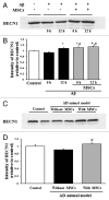
Figure 8. MSCs promote BECN1 expression in vitro and in vivo. Coculture with MSCs in Aβ-treated SH-SY5Y cells exhibited significantly increased BECN1 expression at 6 and 12 h compared with Aβ-treated cells (A and B). Additionally, MSC administration in Aβ-treated mice led to a significant increase in BECN1 expression compared with Aβ-treated mice (C and D). Values are expressed as mean ± SDs, n = 3. *P < 0.05 for Aβ-treated SH-SY5Y cells or mice vs. SH-SY5Y cells or control mice; # P < 0.05 for Aβ-treated SH-SY5Y cells or mice vs. MSCs-treated SH-SY5Y cells or mice.

References

    1. Walsh DM, Selkoe DJ. . Deciphering the molecular basis of memory failure in Alzheimer’s disease. Neuron 2004; 44:181 - 93; http://dx.doi.org/10.1016/j.neuron.2004.09.010; PMID: 15450169 - DOI - PubMed
    1. Sisodia SS, Price DL. . Role of the beta-amyloid protein in Alzheimer’s disease. FASEB J 1995; 9:366 - 70; PMID: 7896005 - PubMed
    1. Haass C, De Strooper B. . The presenilins in Alzheimer’s disease--proteolysis holds the key. Science 1999; 286:916 - 9; http://dx.doi.org/10.1126/science.286.5441.916; PMID: 10542139 - DOI - PubMed
    1. Yankner BA. . Mechanisms of neuronal degeneration in Alzheimer’s disease. Neuron 1996; 16:921 - 32; http://dx.doi.org/10.1016/S0896-6273(00)80115-4; PMID: 8630250 - DOI - PubMed
    1. Mizushima N, Levine B, Cuervo AM, Klionsky DJ. . Autophagy fights disease through cellular self-digestion. Nature 2008; 451:1069 - 75; http://dx.doi.org/10.1038/nature06639; PMID: 18305538 - DOI - PMC - PubMed

Publication types

MeSH terms